filmov
tv
Multiple Sclerosis and MRI Basics: Understand Your Brain Scan

Показать описание
Multiple Sclerosis and the MRI: What is an MRI? How does an MRI work? What is meant by a “MRI with contrast?” These and other important MRI fundamentals are discussed.
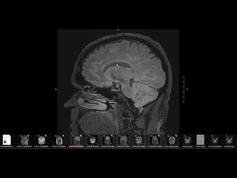
The MRI stands for Magnetic Resonance Imaging and it's the most useful biomarker currently [2018] available to diagnose MS, monitor disease progression, and assess response to disease modifying therapies. Let's discuss the basics:
What is an MRI?
MRI stands for Magnetic Resonance Imaging. The MRI machine is essentially a really big magnet that can take detailed pictures of fat and water. This is fantastic for brain and spinal cord imaging, because of central nervous system is made up of different types of fats and water!
How Does the MRI Work?
The MRI machine uses a giant magnet is used to send out pulses of radio waves. These waves make the protons found in water molecules within the tissues of the brain become “excited” and “stand up at attention.” Then when the radio pulse is turned off, those excited protons “relax” and slowly fall back down. Different neural tissues cause different “relaxation times,” which result different contrasts between the structures of the brain.
Important safety points: The MRI uses a magnet and has NO radiation and NO X-rays
What is an MRI with Contrast?
The contrast dye is called gadolinium. It is injected into your vein partway during MRI. This dye stays in blood stream and lights up blood vessels. The dye can’t cross the blood brain barrier (BBB) so it doesn’t leak out into the brain tissue. If, however a brain with MS experiences new active inflammation, the BBB becomes “irritated” and gets leaky, allowing the contrast dye to leak out. This results in a gadolinium enhancing lesion.
MRI Basics:
-Scanner Strength: measured in units of Tesla. For MS, you want a 1.5T or 3T scanner. NO, a 0.7T open MRI isn’t ok.
-slice thickness: ideally 3mm or thinner.
-Gap between slices: ideally NO gap.
-Sequences for brain: FLAIR SAG, FLAIR AXIAL, T2 AXIAL, T1 PRE CONTRAST AXIAL, T1 POST CONTRAST AXIAL
Please leave your questions and comments below!
Say Howdy on Social:
 0:05:25
Multiple Sclerosis and MRI Basics: Understand Your Brain Scan
 0:10:22
Multiple Sclerosis MRI
 0:01:45
Multiple sclerosis MRI protocol: consensus recommendations and clinician guide
 0:05:11
Multiple Sclerosis and MRI: T1 Black Holes & Brain Atrophy
 0:09:19
Answer Viewers Questions: Multiple Sclerosis MRI
 0:04:10
Multiple Sclerosis and MRI: understanding 'new MRI activity'
 0:03:14
Multiple Sclerosis Vlog: MS MRI
 0:07:54
My Multiple Sclerosis MRI Scan Results (MRI Basics)
 0:47:24
OhioHealth Multiple Sclerosis Lecture-Understanding Your MRI
 0:17:18
Understand your Brain Scan: MS MRI
 0:06:05
Understand Your Scan: Multiple Sclerosis MRI Cervical Spine
 0:00:46
How MRI Can Help Diagnose and Track Multiple Sclerosis
 0:01:57
Magnetic Resonance Imaging in Multiple Sclerosis
 0:02:07
How to identify Multiple Sclerosis & what are its MRI findings? - Dr. Vykunta Raju K N
 0:11:35
Multiple sclerosis – white spots and red flags - part 1 - Making a diagnosis
 0:16:37
Answering Viewers Questions: MS MRI Scan & Multiple Sclerosis Lesions
 0:03:43
Image analysis of the spinal cord in MRI for multiple sclerosis studies
 0:26:29
imaging of Multiple sclerosis
 0:00:16
MS MRI Contrast Dye
 0:05:57
What does MS (multiple sclerosis) look like on brain MRI? Avoid wrongful diagnosis!
 0:05:22
Multiple Sclerosis and the MRI: let's treat brain lesions!
 0:04:07
Diagnosing MS - MRI scan
 0:09:03
Mri MS protocol (Multiple Sclerosis)
 0:10:09
Multiple sclerosis | Types of Multiple Sclerosis | Causes, symptoms, diagnosis, treatment, pathology
Комментарии