filmov
tv
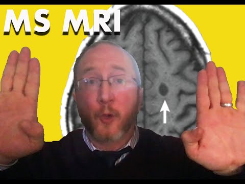
Multiple Sclerosis and MRI: understanding 'new MRI activity'

Показать описание
Multiple Sclerosis and MRI: In this video I answer the question: What exactly does “New MRI activity” mean?
New MS activity occurs one of two ways: either a new clinical attack (e.g. optic neuritis), or new MRI activity. So what exactly does that mean. It means there is a new or enlarged T2 lesions, or a new gad enhancing T1 lesions. OK…so what does THAT mean?
Let’s start by discussing lesions. When talking about MRI scans, the word “lesion” means “a spot.” Seriously, that’s what it means. We see a spot. Then we use other words to describe the lesion, like “T2 lesions” and “gad enhancing lesiosn”
Gad enhancing lesions reveal active CURRENT (ie. As in right now) inflammation in the brain or spinal cord. Lesions tell us that the lesions is new because they only enhance on average for 2-4 weeks (I explain this in a bit more depth in the video).
T2 bright lesions or T2 hyperintense lesions (spots) tell you where there has been inflammation at one time. Maybe it occurred the day after your last MRI, or maybe it’s new today. They are nonspecific to when the lesions occurred (unlike gad enhancing lesions). They typically can be seen for years to come, often for the life that person.
Common locations for MS T2 bright and gad enhancing lesions to show up in MS are:
1. Periventricular: touching the ventricular space in the center of the brain that contains spinal fluid
2. juxta cortical: found right at the junction between the grey matter and the white matter
3. Infratentorial: in the brainstem, or base of the brain (midbrain, pons, medulla, cerebellum)
4. Spinal cord: most commonly in the cervical spinal cord.
MRI is a very powerful tool to survey a given person with MS individual response to a DMT. If a follow up MRI reveals a new gad enhancing lesion, or a new T2 lesions, or an old T2 lesion that has gotten bigger, that’s BAD. And if it occurred despite taking a DMT, then we call that breakthrough disease activity. These findings should trigger a discussion about possible DMT escalation
Please leave your questions and comments below!
Say Howdy on Social:
 0:10:22
Multiple Sclerosis MRI
 0:06:05
Understand Your Scan: Multiple Sclerosis MRI Cervical Spine
 0:04:10
Multiple Sclerosis and MRI: understanding 'new MRI activity'
 0:17:18
Understand your Brain Scan: MS MRI
 0:47:24
OhioHealth Multiple Sclerosis Lecture-Understanding Your MRI
 0:05:25
Multiple Sclerosis and MRI Basics: Understand Your Brain Scan
 0:05:43
Understand Your Scan: MS MRI and Brain Atrophy
 0:09:19
Answer Viewers Questions: Multiple Sclerosis MRI
 0:07:09
Discovered the drug for my aggressive MS in early stage trials
 0:11:32
MS MRI Lesions VS. 'Benign' White Matter Lesions Explained by Neurologist
 0:03:14
Multiple Sclerosis Vlog: MS MRI
 0:05:11
Multiple Sclerosis and MRI: T1 Black Holes & Brain Atrophy
 0:00:46
How MRI Can Help Diagnose and Track Multiple Sclerosis
 0:05:22
Multiple Sclerosis and the MRI: let's treat brain lesions!
 0:11:17
MRIs in Multiple Sclerosis Explained
 0:08:15
Neurologist Reviews MRI Live (Multiple Sclerosis)
 0:06:31
Mayo Clinic Explains Multiple Sclerosis
 0:07:41
How to Diagnose Multiple Sclerosis [In 5 steps]
 0:02:30
What is Multiple Sclerosis? An Overview of MS Causes, Symptoms, Treatments & Research
 0:07:38
Scary Spinal MRI: Multiple Sclerosis Spinal Cord
 0:03:12
Understanding multiple sclerosis and what it does to the body
 0:01:43
Understanding Multiple Sclerosis: The MS Process
 0:17:26
Multiple Sclerosis Diagnostic Criteria [Neurologist Explains]
 0:25:18
Primary Progressive Multiple Sclerosis (PPMS)
Комментарии