filmov
tv
The Pulmonary Hila

Показать описание
 0:09:38
The Pulmonary Hila
 0:01:33
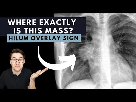
Hilum overlay sign explained
 0:10:28
Hilum of Lung -Structures passing through | Root of Lung | Lung anatomy | [Simplified}
 0:00:32
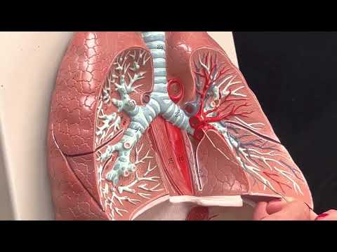
Dr. Benaduce: Respiratory System - Lung hilum
 0:19:23
Hilar Disorders | Chest Radiology Essentials
 0:02:13
the dense hilum sign
 0:02:18
Chest X-Ray guide: how to assess the hilar regions
 0:00:49
Causes of Bilateral Hilar Lymphadenopathy! #pulmonology #internalmedicine #xray
 0:01:06
Chest X-Ray breakdown: assessing the hilar regions
 0:03:46
CXRs, Hilar Lymphadenopathy and High Yield Associations for the USMLE
 0:02:32
Diffferences between the Root of the lung and the Hilum of the lungs
 0:12:08
The Lymph Node Stations in the Chest
 0:08:35
LUNGS PART-2 - ROOT AND RELATIONS - BY DR MITESH DAVE
 0:02:21
Right upper lobe lesion with hilar lymph node
 0:01:41
Root of Lungs/Hilum of Lung : Anatomy 'in 2 minutes'
 0:05:01
Lungs hilum 1
 0:01:13
Hilar lymphadenopathy
 0:02:16
What do lung nodules on a scan mean?
 0:03:49
Mnemonic word 1- SLIPS ;; for DD of bilateral hilar enlargement
 0:08:18
How I Read a Lateral CXR
 0:04:47
Classic Case: Sarcoid
 0:10:30
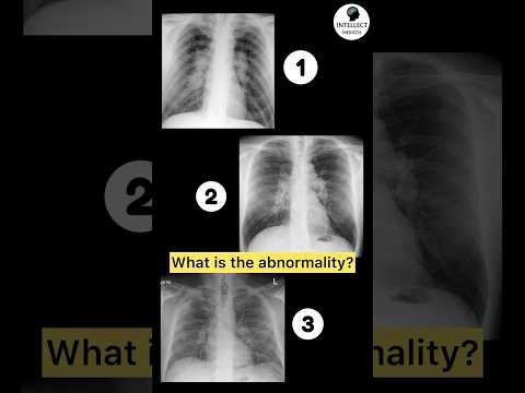
Chest X ray interpretation (in 10 minutes) for beginners🔥🔥🔥 #chestxray #cxr
 0:23:39
Chest X-ray Interpretation | How to Read a CXR | OSCE Guide | UKMLA | CPSA
 0:02:26
What are prominent upper lobe vessels due to?
Комментарии