filmov
tv
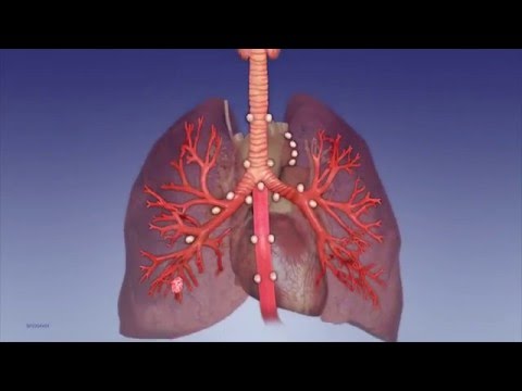
The Lymph Node Stations in the Chest

Показать описание
Check out the IASLC lymph node map here:
Check out the Radiographics article I mentioned here:
Textbooks I like for chest radiology—
Med students and all residents: Felson’s Principles of Chest Roentgenology
Radiology residents: Thoracic Imaging: Pulmonary and Cardiovascular Radiology
Thoracic radiology fellows: Muller’s Imaging of the Chest: Expert Radiology Series
 0:12:08
The Lymph Node Stations in the Chest
 0:03:17
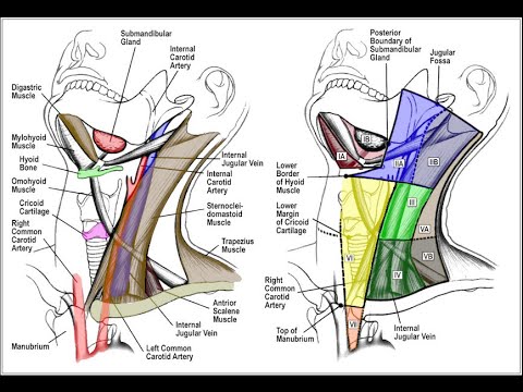
Cervical Lymph Node Stations - a landmark approach
 0:12:40
THORACIC LYMPH NODE STATIONSl Simplified l Updated IASLC Zones & Stations l
 0:05:16
Lymph Node Anatomy-Olympus Bronchoscopy
 0:08:35
Abdominal CT Lymph Node Stations - How to read a CT
 0:07:44
Lymph Nodes Stations of the Chest on CT
 0:13:16
Gastric Lymph Node Stations - Easy to memorize
 0:04:54
Cervical Lymph Node Levels in 5 minutes
 0:00:41
Examination of the lymph nodes of the head and neck
 0:01:32
EBUS Lymph Node Station 2R
 0:10:46
#LYMPH NODE STATIONS IN #LUNGS🫁
 0:18:26
Mediastinal and Hilar Lymph Nodes and the IASLC Lymph Node Map
 0:02:16
CT assessment - exercise on thoracic lymph node stations
 1:49:41
Anatomy of the Abdominal Wall and Abdominal Lymph Node Stations
 0:10:27
Anatomy of the Lymph node | Best Explanation Ever ;)
 0:00:46
Mediastinal lymph node stations
 0:04:29
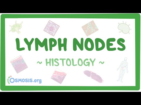
Lymph Nodes: Histology
 0:00:25
Lymph Nodes
 0:13:43
CT: Thoracic Lymph Nodes
 0:38:15
Lymph node map by International Association for the Study of Lung Cancer (IASLC)
 0:52:04
Anatomy of the Thoracic Wall and Thoracic Lymph Node Stations on CT
 0:05:24
How to check your lymph nodes
 0:02:08
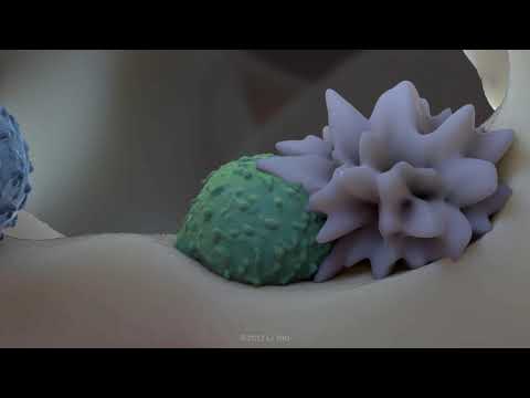
A journey through the lymph node
 0:01:24
EBUS Lymph Node Station 7
Комментарии