filmov
tv
Chest X-Ray guide: how to assess the hilar regions

Показать описание
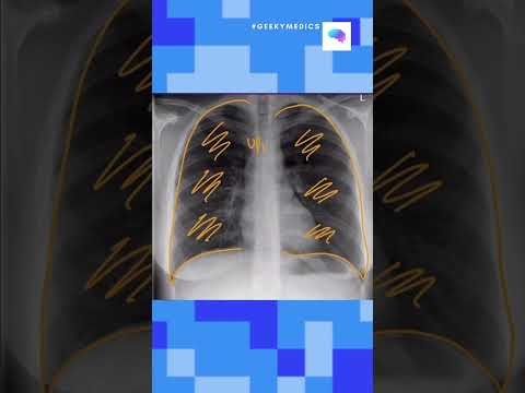
A female in her 50s presents with a few month history of a cough and has a chest X-Ray. What does it show?
————————
LUNG CANCER AND ENLARGED HILAR NODES
👨🏽💻The key to this case is assessing the hilar regions in detail
👨🏽💻The density of the right hilum is abnormal when compared with the left whilst the morphology also does not resemble branching tapering vessels as you would expect
👨🏽💻The second abnormality is an extra contour next to the right hilum. Given this is separate to the hilum this must lie anterior or posterior to the hilum
👨🏽💻Infective consolidation with enlarged nodes is possible but given the lesion is very well defined lung cancer with enlarged nodes at the hilum must be considered
👨🏽💻This was confirmed on CT, PET-CT and CT biopsy
👨🏽💻The key here is to be able to piece apart the hilar regions - remember to assess the density and morphology and look for anything overlying them
FIND THIS USEFUL? SUBSCRIBE FOR THE WHOLE SERIES OF CASE EXPLANATIONS
———-
✅Patient consent obtained
#theradiologist #radiology #radiologist #physician #physicianassistant #medicine #medstudent #medicalstudent #medschool #medicalschool #radtech #xray #medical #chestxray #doctor #medicalstudents #frcr
 0:23:39
Chest X-ray Interpretation | How to Read a CXR | OSCE Guide | UKMLA | CPSA
 0:12:37
Chest X Rays (CXR) Made Easy! - Learn in 10 Minutes!
 0:07:02
Reading a chest X-ray
 0:02:54
Chest X-ray Positioning
 0:03:46
Anatomy of a Chest X-Ray - How to Read a Chest X-Ray (Part 1)
 0:01:00
Chest X-ray Interpretation in 60 seconds - OSCE Guide | UKMLA | CPSA
 0:10:30
Chest X Ray Interpretation Explained Clearly - How to read a chest Xray
 0:12:34
Chest X-rays (CXR) made easy! | COMPLETE GUIDE IN 12 MINUTES
 0:10:48
Dumbbell vs. Kettlebells? Which Gives You FASTER Results?
 0:02:30
A child's guide to hospital: Chest X-Ray
 0:12:51
10 MUST KNOW Chest X-Rays For Medical/PA Finals | CXRs Made Easy
 0:19:44
How to read a chest X-ray (in 20 mins) !
 0:07:19
How to Read a Chest X-ray like a Radiologist! (My Search Pattern)
 0:07:47
How To Read A Chest X-Ray | A Simple Guide On Reading Chest X-Rays | Chest Radiography Lecture Notes
 0:02:18
Chest X-Ray guide: how to assess the hilar regions
 0:44:43
Beginners guide to paediatric chest x-rays
 0:22:35
Beginners Guide to Lines and Tubes - How to Read a Chest Xray
 0:17:57
Basic chest x-ray anatomy
 0:03:54
How to interpret a Chest Xray in under 4 minutes
 0:04:52
How X-Rays Work - How to Read a Chest X-Ray (Part 3) - MEDZCOOL
 0:11:39
HOW TO X-RAY the CHEST in a gurney | AP | Lateral | Portable | radiology program | positioning
 0:04:52
ABCs of Reading a Chest X-ray - How to Read a Chest X-Ray (Part 2) - MEDZCOOL
 0:18:51
How To Read A Chest X-ray
 0:01:56
How To Correctly Rotate A Lateral Chest X-Ray Perfectly – Every Time!!!
Комментарии