filmov
tv
CT Scan - Everything You Need To Know About Computed Tomography

Показать описание
Video Transcript :-
Computed Tomography, also known as CT, is a medical imaging technique that uses X-rays to create detailed cross-sectional images of the body.
CT scans provide a comprehensive view of internal structures, including bones, organs, and soft tissues, by combining multiple X-ray images taken from different angles.
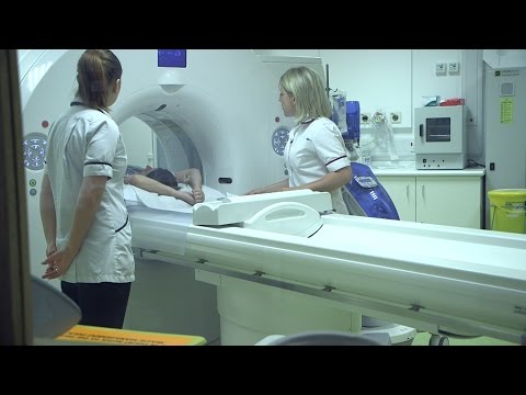
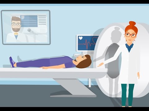
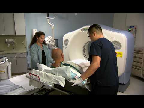
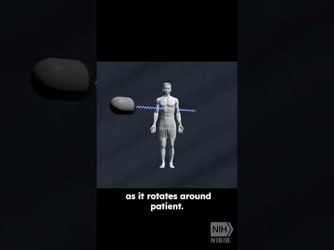
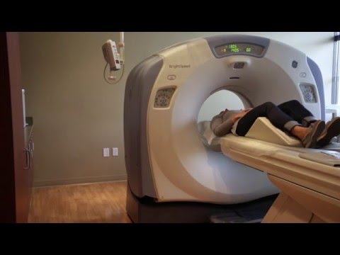
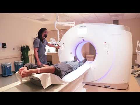
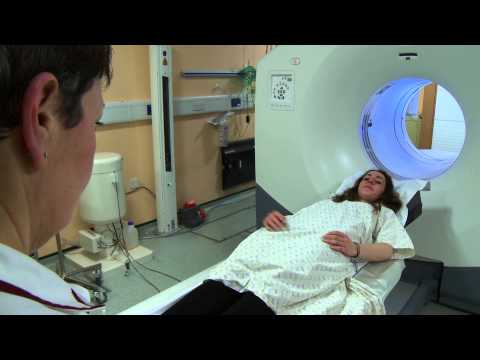
During a CT scan, the patient lies on a table that moves through a large, doughnut-shaped machine called a CT scanner.
Inside the scanner, an X-ray tube rotates around the patient, capturing multiple images from various angles.
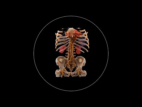
These X-ray images are then processed by a computer to reconstruct a series of cross-sectional images of the body. The images can be displayed in three different planes; axial, coronal, and sagittal, and can be combined to create three-dimensional images.
CT scans are used for a wide range of clinical indications, including the following.
In trauma Assessment, to evaluate injuries to internal organs, bones, and blood vessels.
In cancer Detection and Monitoring to identify tumors, monitor treatment response, and to assess metastasis.
In stroke assessment, to differentiate between bleeding and thrombosis before thrombolytic therapy.
Abdominal and Pelvic Imaging to diagnose conditions such as appendicitis, bowel obstructions, or kidney stones.
And for pre-operative evaluation to provide detailed anatomical information for planning surgical procedures.
CT scans provide high-resolution images with excellent detail of both bone and soft tissues.
They are relatively quick, making them ideal for emergency situations where rapid diagnosis is crucial.
Moreover, CT imaging can be used to visualize a wide range of anatomical areas and conditions.
When comparing CT scans and MRI scans, CT uses X-rays, while MRI uses magnetic fields and radiofrequency waves to generate images.
MRI generally provides better contrast for soft tissues, whereas CT is superior for imaging bone structures.
CT involves ionizing radiation, while MRI does not.
And finally, CT scans are typically faster than MRI scans, which can be beneficial in acute settings.
Since CT scans involve exposure to ionizing radiation, it may increase the risk of cancer over time, especially with repeated scans.
In addition, contrast-enhanced CT scans use contrast agents, which can cause allergic reactions or renal injury in susceptible individuals.
Adverse reactions to contrast media, such as nausea, rash, or more severe reactions, may also occur.
CT scans are generally avoided in pregnant women unless necessary due to the potential risks of radiation to the fetus.
Patients with a history of severe allergic reactions to iodine-based contrast agents may need alternative imaging methods or premedication.
And finally, caution is required with the use of contrast agents in patients with significant kidney dysfunction to avoid further renal complications.
#medicine #ctscan #ctscanner #medtoday #mri #ctvsmri #computedtomography #ctrisks #healtheducation #healthknowledge #medicaleducation #medicalknowledge #science #sciencefacts
Computed Tomography, also known as CT, is a medical imaging technique that uses X-rays to create detailed cross-sectional images of the body.
CT scans provide a comprehensive view of internal structures, including bones, organs, and soft tissues, by combining multiple X-ray images taken from different angles.
During a CT scan, the patient lies on a table that moves through a large, doughnut-shaped machine called a CT scanner.
Inside the scanner, an X-ray tube rotates around the patient, capturing multiple images from various angles.
These X-ray images are then processed by a computer to reconstruct a series of cross-sectional images of the body. The images can be displayed in three different planes; axial, coronal, and sagittal, and can be combined to create three-dimensional images.
CT scans are used for a wide range of clinical indications, including the following.
In trauma Assessment, to evaluate injuries to internal organs, bones, and blood vessels.
In cancer Detection and Monitoring to identify tumors, monitor treatment response, and to assess metastasis.
In stroke assessment, to differentiate between bleeding and thrombosis before thrombolytic therapy.
Abdominal and Pelvic Imaging to diagnose conditions such as appendicitis, bowel obstructions, or kidney stones.
And for pre-operative evaluation to provide detailed anatomical information for planning surgical procedures.
CT scans provide high-resolution images with excellent detail of both bone and soft tissues.
They are relatively quick, making them ideal for emergency situations where rapid diagnosis is crucial.
Moreover, CT imaging can be used to visualize a wide range of anatomical areas and conditions.
When comparing CT scans and MRI scans, CT uses X-rays, while MRI uses magnetic fields and radiofrequency waves to generate images.
MRI generally provides better contrast for soft tissues, whereas CT is superior for imaging bone structures.
CT involves ionizing radiation, while MRI does not.
And finally, CT scans are typically faster than MRI scans, which can be beneficial in acute settings.
Since CT scans involve exposure to ionizing radiation, it may increase the risk of cancer over time, especially with repeated scans.
In addition, contrast-enhanced CT scans use contrast agents, which can cause allergic reactions or renal injury in susceptible individuals.
Adverse reactions to contrast media, such as nausea, rash, or more severe reactions, may also occur.
CT scans are generally avoided in pregnant women unless necessary due to the potential risks of radiation to the fetus.
Patients with a history of severe allergic reactions to iodine-based contrast agents may need alternative imaging methods or premedication.
And finally, caution is required with the use of contrast agents in patients with significant kidney dysfunction to avoid further renal complications.
#medicine #ctscan #ctscanner #medtoday #mri #ctvsmri #computedtomography #ctrisks #healtheducation #healthknowledge #medicaleducation #medicalknowledge #science #sciencefacts
 0:01:50
What is it like to have a CT scan? | Cancer Research UK
 0:01:29
What is a CT scan? | Ohio State Medical Center
 0:03:19
What to Expect During a CT Scan
 0:01:19
What to Expect: How to Prepare for a CT Scan | Bayer
 0:04:16
What is Computed Tomography (CT) and how does it work?
 0:02:37
CT-Scan: What to Expect
 0:03:29
What to Expect: CT Scan | Cedars-Sinai
 0:04:19
What to expect from your CT Scan - a guide for new patients
 0:00:42
Ajj gye mri ki service krne mosam bda pyara tha #mri#ctscan #viral #minivlog #trending #india
 0:00:58
How does a CT scan work?
 0:03:32
What to expect during a CT Scan
 0:02:28
What a Full Body Scan Can Tell You About Your Health
 0:04:45
All About Having A CT Scan - Macmillan Cancer Support
 0:05:15
What to expect during an abdomen and pelvis low-dose CT scan What to expect
 0:01:51
CT Scan (CAT Scan) versus MRI: How They Differ
 0:02:32
What’s the Difference Between an MRI and a CT?
 0:08:09
How a CT scan sees inside of you in 3D
 0:02:52
What is it like to get a CT Scan with Contrast?
 0:04:52
Low Dose CT Scan of the Brain: what to expect at Memorial Healthcare System
 0:00:09
CT Scanner
 0:03:57
So, you're having a CT scan...
 0:05:19
Things To Know Before Undergoing A CT scan
 0:03:53
What is PET/CT and how does it work?
 0:03:13
What is it Like to Have a PET Scan? | Cancer Research UK
Комментарии