filmov
tv
MRI Scan Animation : How magnetic resonance imaging works

Показать описание
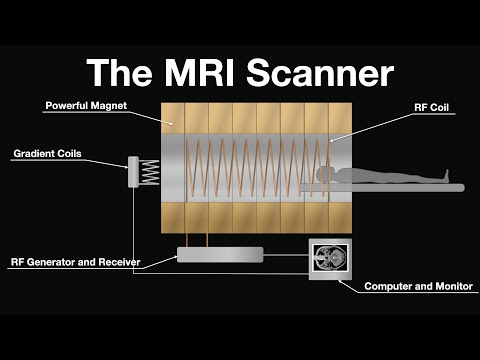
This animation explains how a MRI scan is obtained. It covers how the magnetic resonance signal is produced and detected in the body using powerful magnets and radiofrequency pulses. The role of key components (magnets, gradient coils, and RF coil) are linked to the processes taking place in the body. The animation explains how the MRI signal is localised in image slices of the body using gradient coils. Methods of detecting tissue contrast are introduced, though not explained in detail since the aim of this animation is to introduce the primary concepts of producing an image (proton density, transverse relaxation time, RF pulse sequences and delayed application of localisation fields).
A link to a free quiz on the content of the video is shown at 7:59. Test yourself and check you grasped the main points.
Relevant concepts: nuclear magnetic resonance imaging, protons, precession, Larmor frequency, magnetic field strength, relaxation, phase, transverse and longitudinal vectors, e.m.f., induction and Fourier transformations.
Essential learning for A-Level physics, and medical physics courses at university.
OCR physics A, AQA A-Level physics, Edexcel A-Levrl physics
 0:03:49
How does an MRI work? | MRI basics explained | Animation
 0:07:00
How does an MRI machine work?
 0:03:11
How does an MRI machine work?
 0:08:15
MRI Scan Animation : How magnetic resonance imaging works
 0:01:21
How Does an MRI Scan Work?
 0:17:53
The Insane Engineering of MRI Machines
 0:10:33
MRI Physics | Magnetic Resonance and Spin Echo Sequences - Johns Hopkins Radiology
 0:10:04
How Does An MRI Machine Work Step By Step ?
 0:01:01
#shorts #cartoon #vierl_shorts videos #imaging video #majadar video bird help#funny💯💬🦁🦁🦁🇮🇳...
 0:00:06
MRI 3d animation
 0:03:33
About MRI - Animation
 0:01:00
MRI 3D Medical Animation - Signa by GE Healthcare (3D Animated Magnetic Resonance Imaging Device)
 0:03:05
MRI Scanning for Kids!
 0:00:05
How it feels inside a MRI scanner
 0:03:13
MRI explained: How does it work?
 0:01:01
3D T2 brain MRI imaging
 0:04:37
What happens during an MRI examination?
 0:00:53
Animated Colorized MRI and CT Scans
 0:19:26
What happens behind the scenes of an MRI scan?
 0:03:57
How does an MRI scan work? - in Virtual Reality
 0:02:25
Amerra 3D Medical Animation - MRI-Guided Laser Ablation Technology
 0:04:34
Understanding MRI: What is functional MRI (fMRI)?
 0:00:24
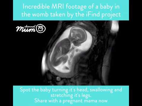
MRI Scan Video of Baby Moving in Womb | Channel Mum
 0:01:17
Magnetic Resonance Imaging (MRI)
Комментарии