filmov
tv
Perforated Vermiform Appendix || Ultrasound || Case 240

Показать описание
Perforated Vermiform Appendix || Ultrasound || Case 240
Clinical Features:
A 17 years old male patient came with
- Right lower abdominal pain
- Vomiting
Ultrasound Features:
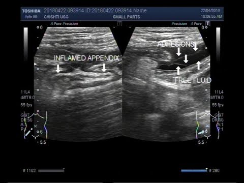
A blind-ended, fluid-filled, distended, non-compressible, tubular structure of intestinal origin, connected to adjacent caecum with poor gut signature, surrounding fat stranding & mild collection is noted at the right iliac fossa with a small wall defect is noted at the tip.
Remember:
Localized collection adjacent to a gangrenous appendix may suggest perforation.
Thank you for watching.
Share with your friends.
Like & Subscribe for more videos.
Don't forget to put your valuable opinions in the Comment box below.
Check the playlist for Congenital Anomaly Lecture videos:
Check the playlist for Imaging Study Lecture videos
Check the playlist for Imaging Study Ultrasound Case videos
Follow us on
#Ultrasound #Radiology #ImagingStudy #Ultrasoundcases #Medical #Imaging #Surgery #aium #doctor #doppler #appendix #appendicitis #radiologist #medicine #isuog
Clinical Features:
A 17 years old male patient came with
- Right lower abdominal pain
- Vomiting
Ultrasound Features:
A blind-ended, fluid-filled, distended, non-compressible, tubular structure of intestinal origin, connected to adjacent caecum with poor gut signature, surrounding fat stranding & mild collection is noted at the right iliac fossa with a small wall defect is noted at the tip.
Remember:
Localized collection adjacent to a gangrenous appendix may suggest perforation.
Thank you for watching.
Share with your friends.
Like & Subscribe for more videos.
Don't forget to put your valuable opinions in the Comment box below.
Check the playlist for Congenital Anomaly Lecture videos:
Check the playlist for Imaging Study Lecture videos
Check the playlist for Imaging Study Ultrasound Case videos
Follow us on
#Ultrasound #Radiology #ImagingStudy #Ultrasoundcases #Medical #Imaging #Surgery #aium #doctor #doppler #appendix #appendicitis #radiologist #medicine #isuog
 0:04:17
Perforated Vermiform Appendix || Ultrasound || Case 240
 0:02:51
Perforated Vermiform Appendix || Ultrasound || Case 110
 0:06:55
How To Scan Appendix | Ultrasound Probe Positioning | Transducer Placement | Abdominal USG Scanning
 0:01:49
Perforated appendicitis
 0:02:08
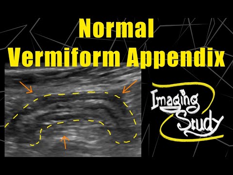
Normal Vermiform Appendix Ultrasound || Ultrasound || Case 116
 0:01:50
Ultrasound Complicated Perforated Appendicitis
 0:02:34
Perforated appendix ultrasound
 0:00:22
Acute Appendicitis 😱🔥😭 Concealed Perforated Appendix #viral #medical #ultrasound #radiology #shorts...
 0:05:12
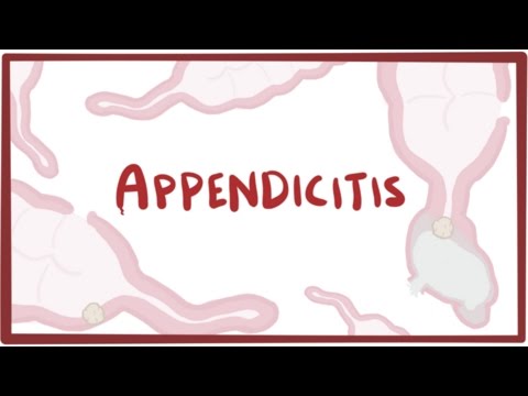
Appendicitis - causes, symptoms, diagnosis, treatment & pathology
 0:02:39
Acute appendicitis perforation ultrasound case
 0:00:17
Appendicitis Ultrasound
 0:00:52
Perforated Appendix. Ultrasound
 0:00:23
Complicated Appendicitis 💯❤️ #medical #ultrasound #infection #health #education #knowledge #shorts...
 0:00:11
When Appendix becomes Infected and Complicated #ultrasound #appendicitis #intestine
 0:00:21
perforated appendix ultrasound by Dr.Haissam Aref,DMS, MSc, MD.Ultrasound
 0:00:13
Ultrasound showing perforated appendix!Appendicitis on ultrasound#youtubeshorts#radiology#ultrasound
 0:06:49
The Appendix on ultrasound and Acute appendicitis | Radiology
 0:01:18
Subacute appendicitis with perforation, Ultrasound and color Doppler video
 0:05:07
Ultrasound Video showing a leakage from the ruptured Appendix.
 0:00:46
Ruptured Appendix | Perforation | Appendicitis | Ultrasound | Right illiac fossa Pain | Tenderness |
 0:01:23
Abdominal Imaging Call Prep Cases: Perforated Appendicitis (CT) Case 1 Discussion
 0:04:37
Appendix Ultrasound Normal Vs Abnormal Image Appearances | Appendicitis USG Scan
 0:01:32
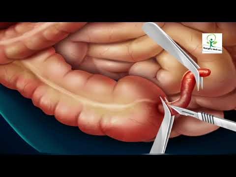
3D Animated Appendix Surgery Explained by//Surgery Medi Care 3D
 0:00:10
ACUTE APPENDICITIS on ULTRASOUND
Комментарии