filmov
tv
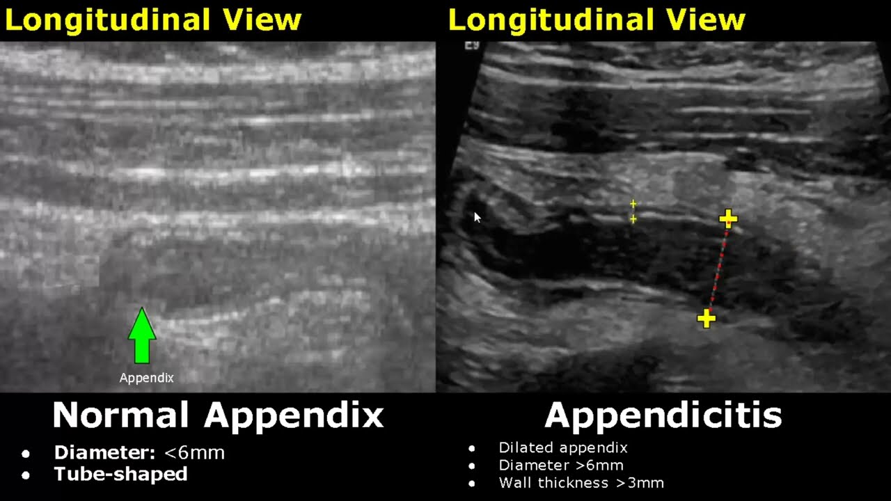
Appendix Ultrasound Normal Vs Abnormal Image Appearances | Appendicitis USG Scan
Показать описание
Appendix Ultrasound Normal Vs Abnormal Image Appearances | Appendicitis USG Scan
Intro - 0:00
Normal Appendix/Appendicitis - 0:07
Appendicitis Color Doppler - 1:05
Appendicitis Spectral Doppler - 1:24
Appendicolith - 1:55
Hyperechoic Periappendiceal Fat - 2:21
Periappendiceal Hyperechoic Structure - 2:49
Periappendiceal Fluid - 3:16
Perforated Appendix - 3:44
You can support our work with a donation:
Guidelines On How To Read And Understand My Videos:
1. Click on the video of your choice.
2. The normal organ images will appear on the left half of the screen.
3. The images with pathologies/diseases will be displayed on the right half of the image.
4. Each case image has description explaining the features of the pathology.
5. Please turn on Subtitles/Closed Caption for easier understanding of the audio, or if you are watching the video on mute.
6. The subtitles may overlap and cover the description. You can move the subtitles away from the description by dragging it with the cursor or with finger touch on smartphone.
Related: images, cases, scans, imaging, pathologies, abnormalities, radiology, sonography, ultrasonography, reporting, hyperechoic, isoechoic, hypoechoic, calcifications, diseases, vermiform
 0:04:37
Appendix Ultrasound Normal Vs Abnormal Image Appearances | Appendicitis USG Scan
 0:03:11
Appendicitis on ultrasound - Radiopaedia's Emergency Radiology Course
 0:01:50
Scanning Technique: Appendix
 0:11:04
US for Suspected Appendicitis in Children
 0:02:52
Scanning the Appendix
 0:06:55
How To Scan Appendix | Ultrasound Probe Positioning | Transducer Placement | Abdominal USG Scanning
 0:06:59
Ultrasound Tutorial: Appendix/Appendicitis | Radiology Nation
 0:17:53
Ultrasound of Acute Appendicitis: Pearls and Pitfalls
 0:01:16
Normal Vs Inflamed Appendix Sonography
 0:05:26
@GEVernova Vscan Appendix Live demo
 0:10:10
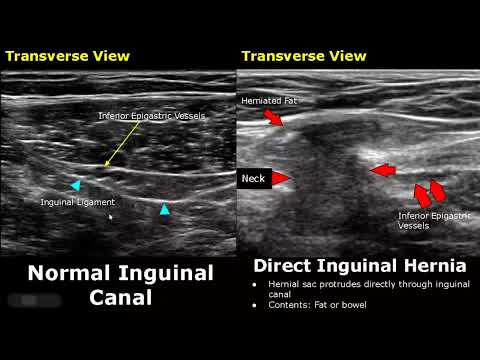
Hernia Ultrasound Normal Vs Abnormal Images | Direct/Indirect Inguinal/Epigastric/Femoral Hernia USG
 1:27:14
Abdominal Ultrasound Normal Vs Abnormal Images | Liver, Gallbladder, Pancreas, Kidney, Hernia USG
 0:02:28
Normal Vermiform Appendix || Ultrasound || Case 50
 0:01:04
Appendicitis: 5 Signs to tell if your Appendix is in risk!
 0:02:05
Appendicitis – Ultrasound Image Interpretation
 0:04:13
Ultrasound Video showing the technique to localize the Inflamed Appendix.
 0:07:30
Pancreas Ultrasound Normal Vs Abnormal Image Appearances Comparison | Pancreatic Pathologies USG
 0:10:02
Gallbladder Ultrasound Normal Vs Abnormal Image Appearances Comparison | Gallbladder Pathologies USG
 0:12:53
Ultrasound of Acute Appendicitis
 0:00:17
Appendicitis Ultrasound
 0:08:31
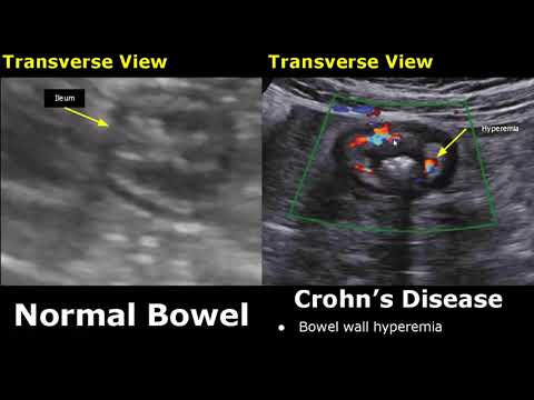
Small Bowel Ultrasound Normal Vs Abnormal Image Appearances | Gastrointestinal Tract (GIT) USG
 0:08:28
The scanning and localization of Acute Appendicitis.
 0:00:23
Complicated Appendicitis 💯❤️ #medical #ultrasound #infection #health #education #knowledge #shorts...
 0:00:46
Normal Appendix Ultrasound
Комментарии