filmov
tv
Macrophages imaging in a live human ovarian cancer tumour slice

Показать описание
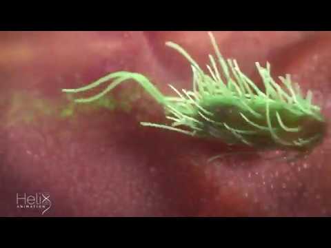
Time-lapse experiment of a live ex vivo human High Grade Serous Ovarian Cancer omentum metastatic tumour slice, stained with CD11b (red), EpCAM (green) and fibronectin (pink).
Images are maximum intensity projections of 8 z slices, acquired every 30 seconds for 30 minutes.
The fibronectin staining shows the extracellular matrix, CD11b shows tumour-associated macrophages (TAMs) and EpCAM shows tumour cells.
This video highlights TAMs' movements within a live human tumour microenvironment.
 0:00:55
 0:00:15
 0:00:16
 0:01:30
 0:01:02
 0:00:59
 0:00:34
 0:01:02
 0:00:47
 0:00:59
 0:00:11
 0:00:23
 0:00:35
 0:00:28
 0:00:14
 0:00:16
 0:00:47
 0:01:01
 0:00:36
 0:05:18
 0:00:21
 0:00:16
 0:01:22
 0:00:42