filmov
tv
Imaging brain tumors - 1 - Introduction and classification

Показать описание
Brain tumors are one of the most common diagnoses addressed in neuroradiology. This covers a wide spectrum of disease, from primary brain tumors like gliomas and glioblastomas to secondary disease like metastases. This lecture covers the spectrum of the most common brain tumors, with an emphasis on primary brain tumor.
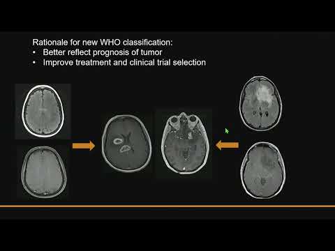
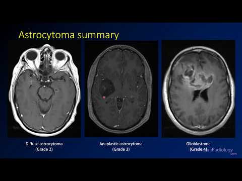
This first video discusses how brain tumors are classified, the genetics of these tumors, and a general approach to brain tumors. Tumors can be divided into a few categories, including astrocytomas, oligodendrogliomas, other low grade glial tumors, and common non-glial tumors. Since the WHO reclassification occurred in 2016, genetic information has been used to classify tumors, with the most important mutations being isocitrate dehydrogenase (IDH), 1p19q codeletion, and MGMT methylation. Oligodendrogliomas must be IDH mutants and 1p19q codeleted, and other tumors are now classified as astrocytomas.
IDH mutation is associated with lower grade astrocytomas and is associated with a survival advantage. Similar, MGMT methylation is associated with a better response to radiation, more pseudoprogression, and better survival.
Additional videos in the playlist will address some of the specific imaging findings and forming a differential diagnosis for brain tumors.
The level of this lecture is appropriate for radiology residents, radiology fellows, and trainees in other specialties who have an interest in neuroradiology or may see patients with brain tumors.
 0:07:51
Imaging brain tumors - 1 - Introduction and classification
 1:33:59
Imaging of brain tumors (part 1): metastases, glioblastoma and beyond...
 0:04:04
Neuroradiology Board Review - Brain Tumors - Case 1
 0:20:15
Imaging Diagnosis of CNS Tumors in Context of the New 2021 WHO Classification
 0:19:05
Brain Tumors
 0:53:10
Imaging Findings of Brain Tumors
 0:36:19
Brain tumour basics - Andrew Dixon (Featured Video)
 1:49:08
Imaging of Brain Tumors - Selected Topics
 1:35:35
ESR/EIBIR Webinar - Data Repositories for AI Evolution in Cancer Diagnosis and Precision Medicine
 0:10:15
Imaging brain tumors - 2 - Astrocytomas
 0:11:27
Emergency Imaging of Brain Tumors: Astrocytomas
 1:36:51
Imaging of Pediatric Brain Tumors
 0:17:45
Advanced Imaging Technologies for Brain Tumors - Javier Villanueva-Meyer, MD
 0:26:05
A Window Into the Brain: Neuro-imaging 101
 1:11:43
Imaging of Pediatric Brain Tumors (part 1): Posterior fossa tumors.
 0:06:48
Imaging brain tumors - 7 - Bonus cases
 1:03:40
Brain Tumor Imaging Standardization Protocols
 0:09:23
Adult Brain Tumors - Radiology Part 1 - RSCP Video Curriculum
 0:36:18
Brain Tumor Imaging Techniques: UCLA Brain Tumor Virtual Conference 2022
 0:08:13
Emergency Imaging of Brain Tumors: Classification
 0:02:00
2-Minute Neuroscience: Brain tumors
 0:02:28
6 Warning Signs of Brain Tumors
 0:26:28
RADIOLOGY OF BRAIN TUMORS - the absolute basics
 0:06:03
Imaging brain tumors - 6 - Common imaging scenarios
Комментарии