filmov
tv
Imaging brain tumors - 2 - Astrocytomas

Показать описание
Brain tumors are one of the most common diagnoses addressed in neuroradiology. This covers a wide spectrum of disease, from primary brain tumors like gliomas and glioblastomas to secondary disease like metastases. This lecture covers the spectrum of the most common brain tumors, with an emphasis on primary brain tumors.
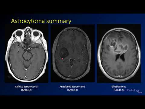
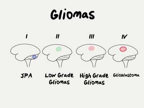
This video discusses imaging and genetic features of astrocytomas, including low grade astrocytomas (WHO grade 2), anaplastic astrocytoma (grade 3), and glioblastoma (grade 4). By definition, these tumors have intact 1p19q. They can have either IDH mutation or IDH wild type, and IDH mutation is associated with increased survival. Higher grade tumors are more likely to have more mass effect, more enhancement, and less well-defined margins.
Additional videos in the playlist will address the imaging findings of other types of brain tumors.
The level of this lecture is appropriate for radiology residents, radiology fellows, and trainees in other specialties who have an interest in neuroradiology or may see patients with brain tumors.
 0:10:15
Imaging brain tumors - 2 - Astrocytomas
 1:33:21
Imaging of brain tumors (part 2): CNS-lymphoma, meningioma, schwannoma and sellar tumors
 0:06:24
Imaging brain tumors - 3 - Oligodendrogliomas
 0:09:31
Emergency Imaging of Brain Tumors: Oligodendrogliomas & Others
 1:33:59
Imaging of brain tumors (part 1): metastases, glioblastoma and beyond...
 0:09:42
Imaging brain tumors - 4 - Other low grade gliomas
 0:02:00
2-Minute Neuroscience: Brain tumors
 0:00:31
Types of brain tumor
 0:02:32
How Radiotherapy in Brain and Spine Tumours – Understanding Neuro-Oncology Radiotherapy
 0:09:37
Emergency Imaging of Brain Tumors: Complications & Summary
 0:07:51
Imaging brain tumors - 1 - Introduction and classification
 0:48:47
Radiology of Brain Tumors: the basics
 0:02:26
Understanding Brain Tumor Survival Rates
 0:21:26
IDoR 2014 - ECR On Demand: Basic 2 'Basic Session on Neuroradiology' (Brain tumours, M.M. ...
 0:01:34
Check Your Health 3D Brain Tumor Imaging
 0:11:27
Emergency Imaging of Brain Tumors: Astrocytomas
 0:20:15
Imaging Diagnosis of CNS Tumors in Context of the New 2021 WHO Classification
 0:06:58
Emergency Imaging of Brain Tumors: Introduction/Role of Imaging
 0:02:28
6 Warning Signs of Brain Tumors
 0:19:45
Comprehensive Brain Tumor Imaging - Soonmee Cha, MD
 0:02:08
Doctor Explains Glioma Brain Tumor
 0:06:03
Imaging brain tumors - 6 - Common imaging scenarios
 0:08:13
Emergency Imaging of Brain Tumors: Classification
 0:01:18
Having Radiotherapy for Brain Cancer
Комментарии