filmov
tv
Parathyroid Hormone (PTH) - Endocrinology

Показать описание
Parathyroid hormone provides a powerful mechanism for controlling extracellular calcium and phosphate concentrations.
Normally there are four parathyroid glands in humans; they are located immediately behind the thyroid gland—one behind each of the upper and each of the lower poles of the thyroid.
The parathyroid gland of the adult human being contains mainly chief cells and a small to moderate number of oxyphil cells. The chief cells are believed to secrete most of the Parathyroid hormone.
Parathyroid hormone secretion is regulated by the plasma calcium concentration. When the total calcium concentration is in the normal range or higher, Parathyroid hormone is secreted at a low level. However, when the plasma calcium concentration decreases , parathyroid hormone secretion is stimulated, reaching maximal rates.
The parathyroid cell membrane contains calcium sensing receptors that are linked to phospholipase C. When the extracellular calcium concentration is increased, calcium binds to the receptor and activates phospholipase C.
Phospholipase C then splits phosphatidylinositol bisphosphate or PIP2 into two molecules diacylglycerol or DAG and inositol triphosphate or IP3.Then IP3 diffuses through the cytoplasm to get to the endoplasmic reticulum, where it binds to a receptor called inositol triphosphate receptor on a ligand-gated calcium channel.
This opens the channel and the calcium that is being stored in the endoplasmic reticulum is released into the cytoplasm, making intracellular calcium levels increase.
High intracellular calcium concentration stops the secretion of Parathyroid hormone.
When extracellular calcium is decreased, there is decreased calcium binding to the receptor, which stimulates Parathyroid hormone secretion.
Parathyroid hormone works to increase extracellular calcium in three ways.First in the bones. In bone, parathyroid hormone receptors are located on osteoblasts which are the bone building cells. Parathyroid hormone binds to receptors on osteoblasts and causes cytokine secretion.
These cytokines then increase the number and activity of the bone resorbing osteoclasts which are bone eating cells.
Osteoclasts breakdown bone, and the two minerals that make up bone - calcium and phosphate, -are released into the blood.
But, in the blood phosphate binds to calciums and form complex calcium which can’t be used cellular processes. To stop this from happening, a second thing that parathyroid hormone will do is bind to receptors on the tubular cells of the kidneys’ proximal convoluted tubules.
This stops the sodium and phosphate cotransporters on the apical surface of the tubular cells from reabsorbing phosphate from the urine.
As a result, phosphate gets lost in the urine, and it’s called phosphaturia.
Parathyroid hormone also binds to principal cells of the distal convoluted tubules.
This causes tubular cells to start making more more sodium calcium channels, which get embedded on their apical surface and increases reabsorption of calcium from urine.
Thirdly, Parathyroid hormone increases calcium absorption of the small intestine.
Parathyroid hormone does not have direct actions on the small intestine, although indirectly it stimulates intestinal calcium absorption via activation of vitamin D.
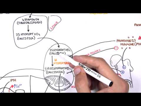
Well, how is vitamin D activated ? Cholecalciferol the precursor of vitamin D is synthesized by keratinocyte cells in the epidermal layer of the skin when exposed to sunlight, but can also come from food. Regardless of the source, cholecalciferol travels to the liver, where the enzyme 25 hydroxylase converts it into 25 hydroxycholecalciferol or calcidiol.
Calcidiol then travels to the proximal tubular cells of the kidneys.
Parathyroid hormone stimulates renal 1 alpha hydroxylase, the enzyme that converts 25 hydroxycholecalciferol to the one 25 dihydroxycholecalciferol or active vitamin D. In turn, active vitamin D stimulates intestinal calcium absorption.
Normally there are four parathyroid glands in humans; they are located immediately behind the thyroid gland—one behind each of the upper and each of the lower poles of the thyroid.
The parathyroid gland of the adult human being contains mainly chief cells and a small to moderate number of oxyphil cells. The chief cells are believed to secrete most of the Parathyroid hormone.
Parathyroid hormone secretion is regulated by the plasma calcium concentration. When the total calcium concentration is in the normal range or higher, Parathyroid hormone is secreted at a low level. However, when the plasma calcium concentration decreases , parathyroid hormone secretion is stimulated, reaching maximal rates.
The parathyroid cell membrane contains calcium sensing receptors that are linked to phospholipase C. When the extracellular calcium concentration is increased, calcium binds to the receptor and activates phospholipase C.
Phospholipase C then splits phosphatidylinositol bisphosphate or PIP2 into two molecules diacylglycerol or DAG and inositol triphosphate or IP3.Then IP3 diffuses through the cytoplasm to get to the endoplasmic reticulum, where it binds to a receptor called inositol triphosphate receptor on a ligand-gated calcium channel.
This opens the channel and the calcium that is being stored in the endoplasmic reticulum is released into the cytoplasm, making intracellular calcium levels increase.
High intracellular calcium concentration stops the secretion of Parathyroid hormone.
When extracellular calcium is decreased, there is decreased calcium binding to the receptor, which stimulates Parathyroid hormone secretion.
Parathyroid hormone works to increase extracellular calcium in three ways.First in the bones. In bone, parathyroid hormone receptors are located on osteoblasts which are the bone building cells. Parathyroid hormone binds to receptors on osteoblasts and causes cytokine secretion.
These cytokines then increase the number and activity of the bone resorbing osteoclasts which are bone eating cells.
Osteoclasts breakdown bone, and the two minerals that make up bone - calcium and phosphate, -are released into the blood.
But, in the blood phosphate binds to calciums and form complex calcium which can’t be used cellular processes. To stop this from happening, a second thing that parathyroid hormone will do is bind to receptors on the tubular cells of the kidneys’ proximal convoluted tubules.
This stops the sodium and phosphate cotransporters on the apical surface of the tubular cells from reabsorbing phosphate from the urine.
As a result, phosphate gets lost in the urine, and it’s called phosphaturia.
Parathyroid hormone also binds to principal cells of the distal convoluted tubules.
This causes tubular cells to start making more more sodium calcium channels, which get embedded on their apical surface and increases reabsorption of calcium from urine.
Thirdly, Parathyroid hormone increases calcium absorption of the small intestine.
Parathyroid hormone does not have direct actions on the small intestine, although indirectly it stimulates intestinal calcium absorption via activation of vitamin D.
Well, how is vitamin D activated ? Cholecalciferol the precursor of vitamin D is synthesized by keratinocyte cells in the epidermal layer of the skin when exposed to sunlight, but can also come from food. Regardless of the source, cholecalciferol travels to the liver, where the enzyme 25 hydroxylase converts it into 25 hydroxycholecalciferol or calcidiol.
Calcidiol then travels to the proximal tubular cells of the kidneys.
Parathyroid hormone stimulates renal 1 alpha hydroxylase, the enzyme that converts 25 hydroxycholecalciferol to the one 25 dihydroxycholecalciferol or active vitamin D. In turn, active vitamin D stimulates intestinal calcium absorption.
 0:09:43
 0:35:03
 0:13:49
 0:02:25
 0:03:57
 0:11:20
 0:12:48
 0:05:55
 0:06:53
 0:01:37
 0:04:57
 0:00:28
 1:04:26
 0:03:15
 0:02:29
 0:01:00
 0:06:46
 0:01:55
 0:01:59
 0:08:01
 0:04:57
 0:03:18
 0:05:47
 0:10:08