filmov
tv
Anatomy Of Eye Part 2 (SCLERA)...... Staphyloma

Показать описание
#mbbs #opthalmology #syndromes
Sclera its parts and the outer membranes.
Anatomy of eye part 1 :-
Anatomy of eye part 2 :-
Anatomy of eye part 3 :-
Anatomy of eye part 4 :-
Anatomy of eye part 5 :-
Anatomy of eye part 6 :-
Sclera its parts and the outer membranes.
Anatomy of eye part 1 :-
Anatomy of eye part 2 :-
Anatomy of eye part 3 :-
Anatomy of eye part 4 :-
Anatomy of eye part 5 :-
Anatomy of eye part 6 :-
 0:06:35
GCSE Biology - How the Eye Works (Part 2) - Accommodation #32
 0:02:49
EYE ANATOMY IN 3 MINUTES!
 0:36:14
Anatomy of the Orbit: Part 2: III, IV & VI Nerves and Ophthalmic Vessels, Dr Adel Bondok
 0:06:53
Professor Long - Eye Anatomy 2, Internal Anatomy
 0:12:19
Anatomy of Eye Part 2 Vascular Layer of Eye
 0:45:46
Special Senses | Eye Anatomy
 0:10:46
Anatomy of the Eye Part 2
 0:01:45
Human A&P: Anatomy of the Eye
 1:03:35
Our Grandchildren Will Look Like Two Peas in a Pod
 0:09:55
Eyeball Anatomy
 0:05:09
GCSE Biology - How the Eye Works (Part 1) - Structure of the Eye & Iris Reflex #31
 0:17:05
Basic Eye Anatomy and Physiology
 0:07:20
Eye Anatomy and Function - Made Easy
 0:03:04
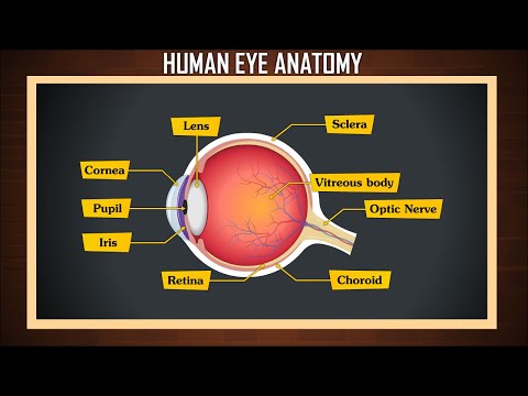
Human Eye Anatomy | Structure and function | Parts of the Eye
 0:10:09
Eye Anatomy | Review and Practice
 0:10:02
Professor Long - Eye Anatomy 1, External Anatomy and Lacrimal Structure
 0:10:22
Parts of the eye | Human eye & the colourful world | Khan Academy
 0:04:42
Human Anatomy Eye
 0:51:01
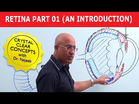
Retina | An Introduction | Part 1
 0:26:40
Aqueous Humor | Production, Circulation & Drainage part 2/2
 0:03:51
Causes of Red Eye - Part 2: ANTERIOR UVEITIS (Iritis)
 0:10:35
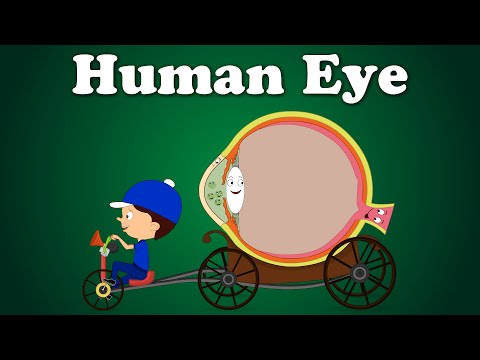
Human Eye | #aumsum #kids #science #education #children
 0:45:42
Lecture 1: Anatomy of the eyeball (Part 2), Anatomy of ocular structures
 0:04:17
Human Eye - The Dr. Binocs Show | Best Learning Videos For Kids | Peekaboo Kidz
Комментарии