filmov
tv
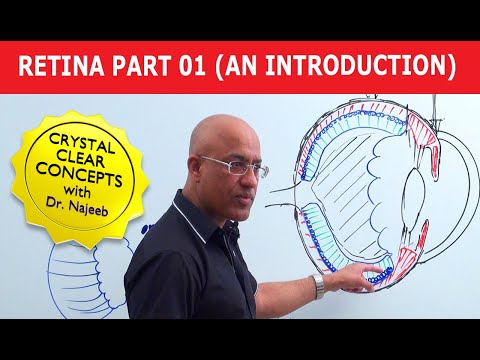
Retina | An Introduction | Part 1

Показать описание
Retina | An Introduction | Part 1
Like this video?
---------------------------------------------------------------------------------------------------------------------------
Why sign up for premium membership? Here's why!
Membership Features for premium website members.
1. More than 800+ Medical Lectures.
2. Basic Medical Sciences & Clinical Medicine.
3. Mobile-friendly interface with android and iOS apps.
4. English subtitles and new videos every week.
5. Download option for offline video playback.
6. Fanatic customer support and that's 24/7.
7. Fast video playback option to learn faster.
8. Trusted by over 2M+ students in 190 countries.
---------------------------------------------------------------------------------------------------------------------------
▬▬▬▬▬▬▬▬▬▬ Contents of this video ▬▬▬▬▬▬▬▬▬▬
(0:00-3:40)
Delicate membranous light-sensitive part of the eye, location in the eye ball;
Outermost protective layer= Sclera & Cornea.
Middle layer (Uveal Tract) = Choroid, Ciliary body & Iris.
Innermost layer= Retina; pigmented epithelium, Innermost Neuronal layer of Retina.
(3:41-9:30)
Exact location of Retina; sandwiched between Vitreous Body inside (Vitreous aspect of retina) and Choroid outside (Choroidal part of retina).
Ora Serrata; Wavy margins where light sensitive part of retina terminates anteriorly.
(9:35-13:20)
Through the Ophthalmoscope: Only posterior part of retina i.e., Optic Disc and part around it visible; Ocular Fundus.
Non-light sensitive part of retina; continuing beyond Ora Serrata.
(13:22-23:20)
Embryological Development of Eye: CNS development; Neural Tube; Optic Vesicle to Optic Cup; Outer and Inner layers differentiate into outer pigmented & inner neuronal retinal layers.
Retina, therefore is the only part of CNS visible through ophthalmoscope.
(23:25-29:10)
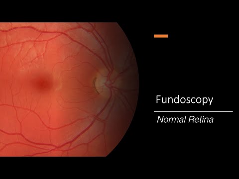
Fundoscopy: Posterior pole of fundus(yellowish) [Macula Lutea]; Optic Disc nasally/medially (pale colored).
Macula Lutea in detail: Has undefined margins, slight depressed area in center; Fovea Centralis.
(29:15-34:25)
Optic Disc in detail: Highly vascular (Central Retinal Artery coming out of it), Optic Disc; point of Retina from where all optic nerve fibers are moving out of retina through lamina cribrosa as Optic Nerve.
Technically Optic Nerve is a CNS tract and not a nerve per say.
(34:30-38:40)
Optic Disc continued: Central Retinal Artery, branches after passing through the Optic Disc & its Nasal & Temporal branches then supply inner parts of Retina.
Outer part of Retina supplied by Choriocapillaris.
(38:41-41:30)
Optic Disc continued; Central Retinal Artery (CRA) continued: Fovea Centralis (FC); the area of maximal visual acuity, depends only on Choriocapillaris for nutrition & not CRA, therefore NO Blood Vessels: Avascular.
Further comparison of Macula Lutea and Optic Disc.
(41:31-43:44)
Clinical Co-relate of Optic Disc (OD): Physiological Cup: Depression in OD; In Glaucoma when ICP is very high, this cup is pushed backwards; pathologically cupped/depressed. No rods & cones here so; Blind Spot.
Comparison continued: In Fovea Centralis (FC), cones are packed very tightly & blood vessels basically pushed aside to reveal FC; hence thinnest area of the retina; sharpest acuity.
(43:48-45:30)
Summary of comparison b/w OD & Macula Lutea (ML); 1- OD nasally situated, ML central but inferolateral to OD.
2-OD = 1.5mm, while ML = 5-6mm.
3-OD is pale, ML yellowish.
4- OD = Blood vessels coming in & out; highly vascular, while ML = Avascular.
5- OD is a blind spot while ML has point of maximal visual acuity.
6-OD has 'Physiological cupping' while ML has Fovea Centralis.
7-OD has sharper margins, while ML much diffused, undefined margins.
(45:31-51:00)
Blood supply of Fovea Centralis. Descriptive schematic diagram of OD, ML, Optic Nerves and Retinal Arteries, along with their positions in eyeball from above.
---------------------------------------------------------------------------------------------------------------------------
Join this channel to get access to the perks:
Like this video?
---------------------------------------------------------------------------------------------------------------------------
Why sign up for premium membership? Here's why!
Membership Features for premium website members.
1. More than 800+ Medical Lectures.
2. Basic Medical Sciences & Clinical Medicine.
3. Mobile-friendly interface with android and iOS apps.
4. English subtitles and new videos every week.
5. Download option for offline video playback.
6. Fanatic customer support and that's 24/7.
7. Fast video playback option to learn faster.
8. Trusted by over 2M+ students in 190 countries.
---------------------------------------------------------------------------------------------------------------------------
▬▬▬▬▬▬▬▬▬▬ Contents of this video ▬▬▬▬▬▬▬▬▬▬
(0:00-3:40)
Delicate membranous light-sensitive part of the eye, location in the eye ball;
Outermost protective layer= Sclera & Cornea.
Middle layer (Uveal Tract) = Choroid, Ciliary body & Iris.
Innermost layer= Retina; pigmented epithelium, Innermost Neuronal layer of Retina.
(3:41-9:30)
Exact location of Retina; sandwiched between Vitreous Body inside (Vitreous aspect of retina) and Choroid outside (Choroidal part of retina).
Ora Serrata; Wavy margins where light sensitive part of retina terminates anteriorly.
(9:35-13:20)
Through the Ophthalmoscope: Only posterior part of retina i.e., Optic Disc and part around it visible; Ocular Fundus.
Non-light sensitive part of retina; continuing beyond Ora Serrata.
(13:22-23:20)
Embryological Development of Eye: CNS development; Neural Tube; Optic Vesicle to Optic Cup; Outer and Inner layers differentiate into outer pigmented & inner neuronal retinal layers.
Retina, therefore is the only part of CNS visible through ophthalmoscope.
(23:25-29:10)
Fundoscopy: Posterior pole of fundus(yellowish) [Macula Lutea]; Optic Disc nasally/medially (pale colored).
Macula Lutea in detail: Has undefined margins, slight depressed area in center; Fovea Centralis.
(29:15-34:25)
Optic Disc in detail: Highly vascular (Central Retinal Artery coming out of it), Optic Disc; point of Retina from where all optic nerve fibers are moving out of retina through lamina cribrosa as Optic Nerve.
Technically Optic Nerve is a CNS tract and not a nerve per say.
(34:30-38:40)
Optic Disc continued: Central Retinal Artery, branches after passing through the Optic Disc & its Nasal & Temporal branches then supply inner parts of Retina.
Outer part of Retina supplied by Choriocapillaris.
(38:41-41:30)
Optic Disc continued; Central Retinal Artery (CRA) continued: Fovea Centralis (FC); the area of maximal visual acuity, depends only on Choriocapillaris for nutrition & not CRA, therefore NO Blood Vessels: Avascular.
Further comparison of Macula Lutea and Optic Disc.
(41:31-43:44)
Clinical Co-relate of Optic Disc (OD): Physiological Cup: Depression in OD; In Glaucoma when ICP is very high, this cup is pushed backwards; pathologically cupped/depressed. No rods & cones here so; Blind Spot.
Comparison continued: In Fovea Centralis (FC), cones are packed very tightly & blood vessels basically pushed aside to reveal FC; hence thinnest area of the retina; sharpest acuity.
(43:48-45:30)
Summary of comparison b/w OD & Macula Lutea (ML); 1- OD nasally situated, ML central but inferolateral to OD.
2-OD = 1.5mm, while ML = 5-6mm.
3-OD is pale, ML yellowish.
4- OD = Blood vessels coming in & out; highly vascular, while ML = Avascular.
5- OD is a blind spot while ML has point of maximal visual acuity.
6-OD has 'Physiological cupping' while ML has Fovea Centralis.
7-OD has sharper margins, while ML much diffused, undefined margins.
(45:31-51:00)
Blood supply of Fovea Centralis. Descriptive schematic diagram of OD, ML, Optic Nerves and Retinal Arteries, along with their positions in eyeball from above.
---------------------------------------------------------------------------------------------------------------------------
Join this channel to get access to the perks:
 0:51:01
Retina | An Introduction | Part 1
 0:07:22
Introduction to the Retina
 0:21:17
An Eye on Education: Introduction to the Eye, Retina & Vision
 0:00:26
What is the Retina?
 0:09:55
Eyeball Anatomy
 0:00:45
Easy Way To Remember Layers Of Retina
 0:00:06
Retina ophthalmology
 1:34:36
Episode 1 - Introduction to Retina
 1:14:39
THE X FILES | Retina | EP 01| KSOS
 0:07:24
Introduction of retina
 0:17:05
Basic Eye Anatomy and Physiology
 0:00:48
Retina Model
 0:00:53
Brown Retina Institute Practice Introduction
 0:01:52
Healthy Retina: Fundoscopy
 0:20:53
Introduction to Cognitive Neuroscience: Session 4.1 (Neural basis of visual processing: retina)
 0:00:35
The human eye . #cornea #retina
 0:03:07
The new MacBook Pro - Design, Performance and Features - Apple
 0:03:56
Layers of retina mnemonic
 0:02:20
Anatomy of Retina - Introduction / what is Retina and its function
 0:00:31
Easy way to remember Layers of Retina
 0:23:41
Ophthalmology Made Ridiculously Easy | 1st Edition | Digital Book
 0:03:36
Inverted Retina
 0:00:23
BIDMC Retina Service Introduction
 0:05:45
Introducing The MacBook Pro with Retina display
Комментарии