filmov
tv
Electrical Cardiac Conduction System of the Heart EKG ECG Animation Nursing School NCLEX-RN
Показать описание
Electrical Cardiac Conduction System of the Heart and how it corresponds to the EKG
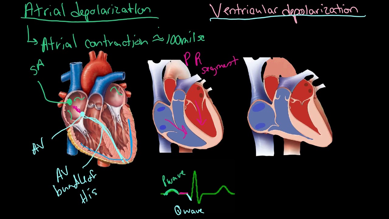
Step 1: The SA node sends impulse through atria causing atrial depolarization and this leads to atrial contraction. This is shown by the P wave.
Step 1.5: The atria contract pushing blood into the ventricles. Meanwhile, the SA node sends an electrical impulse to the AV node. This is shown by the PR interval.
Step 2: The AV node sends impulse through the intraventricular septum of the heart to the apex of the heart along the (Step 4) Bundle of His/ AV Bundle starting ventricular depolarization This is shown by the Q wave.
Step 4: The electrical impulse branches off into the Left Bundle Branch and the Right Bundle Branch and the to the (Step 5) Purkinje Fibers continuing ventricular depolarization. This is shown by the R wave. The ventricles complete depolarization and this is shown by the S wave.
Step 5.5: The ventricles contract pushing blood into the body and lungs. This is shown by the ST segment.
Finally the ventricles repolarize and this is shown by the T wave.
The EKG used in this video is a normal sinus rhythm. For information about abnormal rhythm or arrhythmias keep an eye out for future videos. Please subscribe below to keep watching, learning, and tRNsforming!
#nclex #heart #rn #cardiacconduction #ekg
 0:16:17
 0:03:17
 0:03:02
 0:48:01
 0:05:58
 0:15:59
 0:01:38
 0:23:27
 0:15:55
 0:03:56
 0:17:46
 0:18:09
 0:00:33
 0:09:28
 0:20:33
 0:09:19
 0:01:51
 0:04:48
 0:04:24
 0:01:42
 0:04:04
 0:03:15
 0:00:56
 0:11:30