filmov
tv
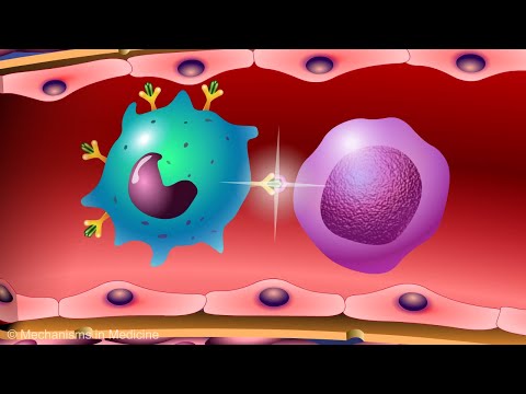
How to Characterize Immune Responses With Flow Cytometry

Показать описание
Combining analysis of surface markers and intracellular signaling readouts through multiplexed flow cytometry adds power to your experiments. However, this can present challenges when designing antibody panels. This video walks through an example experiment to illustrate how antibodies can be selected for a panel to assess activation of T cell subpopulations.
Transcript:
If you are faced with the challenge of designing antibody panels to analyze intracellular signaling with flow cytometry, and you’re not sure how to find your way, that’s okay.
You can do it. We can help.
We will walk through an example experiment to illustrate how you can build an antibody panel for multiplexed flow cytometry to assess immune cell activation.
Multiplexing adds power to your flow cytometry analysis. Phenotypic surface markers pinpoint which cell populations exhibit responses to treatment or experimental perturbation, while intracellular readouts probe the signaling pathways involved in that response.
In our example experiment, our goal is to assess activation of T cell lymphocytes within a heterogeneous PBMC sample. Start building your panel by selecting appropriate surface markers so that you can gate populations and subpopulations in your analysis. In our example, we used CD3 as a pan-T cell marker and CD4 as a helper T cell marker.
Add one or more intracellular targets as readouts for signaling pathway activation, protein expression, or other biological functions. We used a phospho-specific antibody to detect phosphorylation of SLP-76 as a readout of T cell activation. Finally, consider incorporating a live-dead viability stain such as a Ghost Dye in your panel to allow exclusion of dead cells from analysis.
Here are some best practices to ensure your flow cytometry results are reliable and reproducible. When building your panel, make sure to select antibodies or antibody conjugates that have been validated in flow cytometry. Performance in other applications does not tell you how that antibody will perform in flow.
It’s also important to check that all of the antibodies in your panel are compatible with the fixation and permeabilization conditions used in your protocol. Testing and optimizing of protocol steps may be required if this information isn’t available. Finally, when selecting fluorophores, avoid spectral overlap to the extent possible, or, incorporate fluorescence compensation into your analysis when necessary.
To prepare our live PBMCs for flow cytometry analysis, we first incubated cells with Ghost Dye and washed. Then, we activated the T cells by treatment with antibodies to induce crosslinking of CD3 and CD28 for 15 minutes prior to fixation, permeabilization, and labeling with antibodies for analysis.
To analyze SLP76 activity in T cells, we employed a sequential gating scheme. Forward and side scatter were used in Gate 1 to select lymphocytes. The live/dead selection with Ghost Dye was used for Gate 2, followed by selecting for CD3 expression in Gate 3 and CD4 expression in gate 4. Gating in this way enables you to quickly zero in on relevant cell populations and get to the data that matters.
In our experiment, T cell activation is clearly distinguishable in CD3/CD4 positive T cells, and the percentage of cells positive for phospho SLP-76 expression after activation is higher in the gated subpopulation than in the bulk PBMC’s.
© 2019 Cell Signaling Technology. All rights reserved. Cell Signaling Technology, CST, and XP are trademarks of Cell Signaling Technology. All other trademarks are the property of their respective owners.
 0:04:00
How to Characterize Immune Responses With Flow Cytometry
 0:08:56
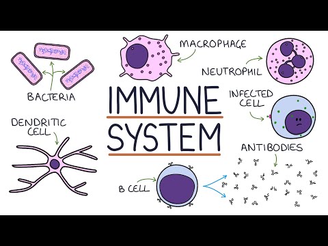
Immune System
 0:16:31
Introduction to the immune system
 0:15:17
Understanding the Immune System in One Video
 0:06:53
Immune Response Explained: T-Cell Activation
 0:05:13
Introduction to Innate Immunity
 0:10:48
How The Immune System ACTUALLY Works – IMMUNE
 0:32:38
Deep Characterization of Viral and Self-Antigen Peptide-Induced Cellular Immune Responses
 0:03:03
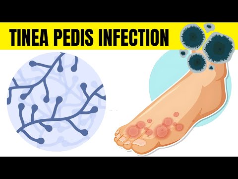
Tinea Pedis Infection (Athlete's Foot) - Causes, Types, Signs & Symptoms, And Treatment
 0:07:01
Immune System: Innate and Adaptive Immunity Explained
 0:09:13
Immune System, Part 1: Crash Course Anatomy & Physiology #45
 0:01:34
News Method to Characterize Immune Cells in Tumors
 0:14:21
Immunology | Immune System: Overview
 0:25:09
IMMUNE SYSTEM MADE EASY- IMMUNOLOGY INNATE AND ADAPTIVE IMMUNITY SIMPLE ANIMATION
 0:03:46
Humoral and Cell Mediated Immunity
 1:01:42
Cell-based Assays to Functionally Characterize Immunotherapy and the Immune Response to Viral...
 0:04:46
GCSE Biology - Immune System (Defences Against Pathogens) #38
 0:25:05
immunology; immune system - adaptive and innate immunity
 0:13:44
The Immune System: Innate Defenses and Adaptive Defenses
 0:01:12
How Does the Immune System Respond to a Pathogen
 0:41:48
Immunology | Adaptive Immunity
 0:03:37
Vaccines and the Immune Response: How Vaccines Work
 0:06:08
Primary and secondary Immune response
 0:18:46
Lecture 1c: Categories of Immune Responses
Комментарии