filmov
tv
MRI / 3D Brain Anatomy: Internal Capsule, Thalamus & Brainstem

Показать описание
The first in a series using MRI next to my 3D models, aiming to simplify the 3D comprehension of the anatomy of the brain.
MRI citation: Puccio et al.
--
In this video, we’re going to compare MRI images of the brain to this schematic 3D model.
By doing so, we’ll get a better intuition for how the parts of the brain fit together.
The three sets of images you see here are taken in the coronal, sagittal and transverse planes.
This corresponds to these planes on our 3D model - coronal, sagittal and transverse.
Throughout the video, I’ll be moving through all three planes.
Notice how, if I click and drag on this image, we move through the slices on the other planes.
Take a look at the coronal plane, as I drag the cursor through the sagittal plane, from right to left.
We’ll bring that back to the centre. Now look at the transverse plane images, as I drag the cursor from the top of the brain downward. This is also known as the axial plane.
Occasionally I’ll enlarge one plane like so. This is a sagittal plane image, in the midline, so we are looking at the very middle of the brain here.
Now let’s look at our 3D model for a moment.
To put it in context, we have this 2D body outline, but let’s remove that now.
To really ground ourselves, let’s recreate the image on the left. So we’ll take away half of all the models. And then we can line up our sagittal image.
Ok, now that you have a sense of perspective, let’s begin talking about the anatomy.
The first thing we see is the cortex - the grey matter on the outside of the brain.
Looking over to our MRI now, see the darker grey cortex here. It’s all bumpy and knobbly. Which is why it goes by the name cortex, meaning bark in Latin, because anatomists from back in the day thought it felt like the bark of a tree.
The cortex is divided into lobes, which we’ll just go through briefly, that’s the frontal, parietal, occipital and temporal lobes.
Beneath the grey matter, moving over to look at our MRI again, is all this white matter.
White matter is the axons, or the electrical wires, of the neurons that make up the brain.
The cell body, the part of the neuron that generates or manipulates information, is in the cortex.
Now, in order to move our muscles a message has to be generated in the brain and sent down the spinal cord.
Let’s think about the passage of that information in the brain.
So the cell body in the motor cortex, which sits in the back part of the frontal lobe, generates the message.
It sends it down the axon, part of the white matter here. The axon passes through this area, called the internal capsule.
 0:06:19
MRI / 3D Brain Anatomy: Hypothalamus, Pituitary Gland, Fornix, Corpus Callosum & 3rd Ventricle
MRI / 3D Brain Anatomy: Internal Capsule, Thalamus & Brainstem
 0:15:05
Ventricles and Cisterns of the Brain | Radiology anatomy part 1 prep | MRI brain
 0:03:22
Normal Brain MRI Anatomy - Neuroradiology Made simple
 0:11:58
Basic Parts of the Brain - Part 1 - 3D Anatomy Tutorial
 0:20:11
Cranial Nerve Anatomy on MRI
 0:00:11
🧠 The Brain in Detail: Exploded View Anatomy 🧠 #anatomy
 0:23:53
Cerebral Vascular Anatomy And Imaging
 0:06:12
Basal Ganglia 3D Tour
 0:00:41
Brain MRI 🧲 🧠 #mri #radiology
 0:00:58
3D View Of Dissected Brain
 0:00:15
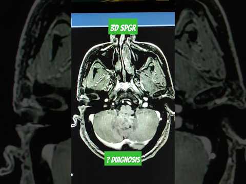
3D SPGR Post Contrast Brain Mri #viral #mri #shorts
 0:00:20
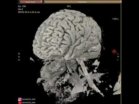
3D VRT MRI of brain and face
 0:21:14
Neuroanatomy on MRI | Part 1| Cerebrum, Basal Ganglia, Thalamus, Internal Capsule & Lesions
 0:08:18
How to Read a Brain MRI: Basic Search Pattern & Sequences Explained
 0:09:25
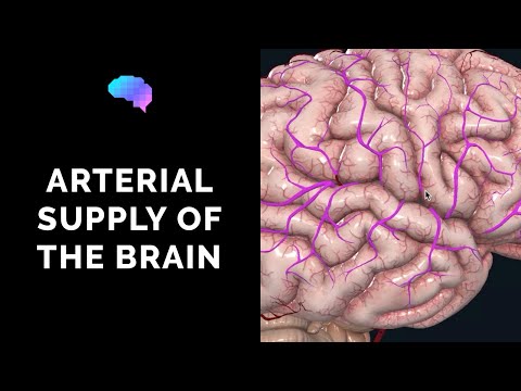
Blood Supply to the Brain (3D Anatomy Tutorial) | UKMLA | CPSA | PLAB 2
 0:00:12
Head and Brain Internal Structure in 3D animation 😱 #brain #animation #medical
 0:01:00
3D Interactive Animation Reveals Hemorrhaging Found In MRI Scans of Brain
 0:09:35
Brain MRI scan protocols, positioning and planning
 0:08:12
MRI Brain Anatomy and Physiology in English
 0:00:16
3 workhorse Brain MRI sequences! #shorts #radiology #medschool
 0:01:14
3D Brain for MRI Hippocampus.
 0:04:01
Brain Anatomy On an MRI (Magnetic Resonance Imaging) Scan
 0:00:11
CT Angiography of Head and Neck Anatomy #angiography #neckangio #ctscan #radiology #angiography
Комментарии