filmov
tv
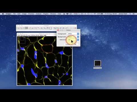
Counting Cells with ImageJ
Показать описание
Keep in mind that ImageJ processing is not perfect. However, the error of missing cells may be "corrected" by the error of including some noise.
Steps in Video:
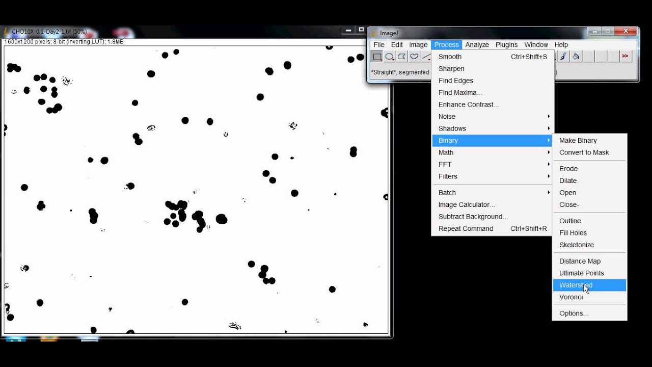
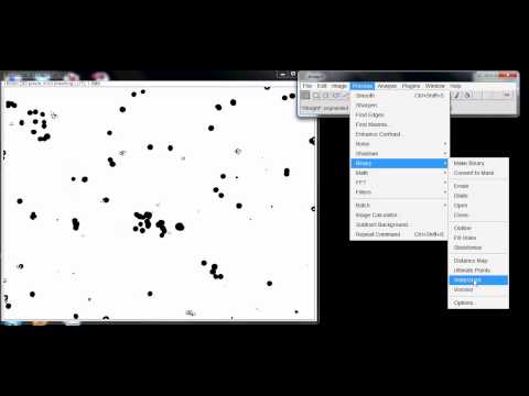
Process - Subtract Background
Image - Adjust - Threshold
Process - Binary - Fill Holes
Process - Binary - Convert to Mask
Process - Binary - Watershed
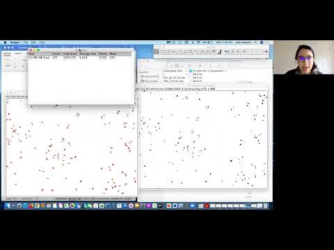
Analyze - Analyze Particles
Basic description of tools:
Subtract Background - cancel out noise of the background
Threshold - change the image to binary image of red, black and white, or blue
Fill Holes - fill empty spaces between rings to make circles
Convert to Mask - allows for subsequent processing
Watershed - automated separation of separate fused cells by a 1 pixel line
Analyze Particles - process the image to acquire a cell count
Size - parameter of what cells to include in data by area (pixels^2)
Circularity - parameter of what cells to include by how close to a circle the shape appears. The maximum bound (1.0) means a perfect circle. The minimum bound deviates from being a circle.
Show - Outlines will trace the outside of the mask images. Masks will show the black and white image similar during thresholding.
Exclude Edges - ImageJ will not include cells that are not fully contained in the boundaries of the image
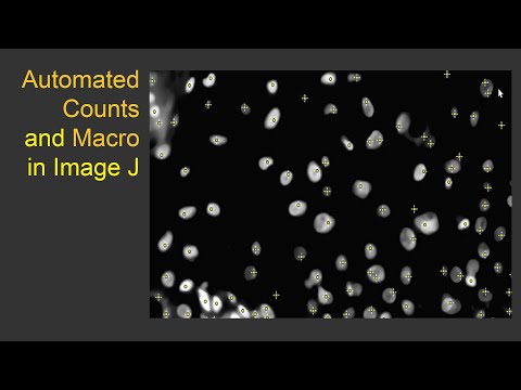
Macro Code:
run("Subtract Background...", "rolling=12");
setAutoThreshold("Default dark");
setThreshold(35, 255);
run("Convert to Mask");
run("Fill Holes");
run("Convert to Mask");
run("Watershed");
run("Analyze Particles...", "size=120-Infinity circularity=0.00-1.00 show=Outlines display exclude clear summarize");
Steps in Video:
Process - Subtract Background
Image - Adjust - Threshold
Process - Binary - Fill Holes
Process - Binary - Convert to Mask
Process - Binary - Watershed
Analyze - Analyze Particles
Basic description of tools:
Subtract Background - cancel out noise of the background
Threshold - change the image to binary image of red, black and white, or blue
Fill Holes - fill empty spaces between rings to make circles
Convert to Mask - allows for subsequent processing
Watershed - automated separation of separate fused cells by a 1 pixel line
Analyze Particles - process the image to acquire a cell count
Size - parameter of what cells to include in data by area (pixels^2)
Circularity - parameter of what cells to include by how close to a circle the shape appears. The maximum bound (1.0) means a perfect circle. The minimum bound deviates from being a circle.
Show - Outlines will trace the outside of the mask images. Masks will show the black and white image similar during thresholding.
Exclude Edges - ImageJ will not include cells that are not fully contained in the boundaries of the image
Macro Code:
run("Subtract Background...", "rolling=12");
setAutoThreshold("Default dark");
setThreshold(35, 255);
run("Convert to Mask");
run("Fill Holes");
run("Convert to Mask");
run("Watershed");
run("Analyze Particles...", "size=120-Infinity circularity=0.00-1.00 show=Outlines display exclude clear summarize");
 0:05:06
Counting Cells with ImageJ
 0:01:07
How to count cells using ImageJ
 0:06:11
How to Count Cells Using ImageJ | How to Count Cells in Imagej | Imagej Cell Counting |
 0:06:13
Best method of cell counting using image J (Fiji)
 0:06:56
How to use ImageJ software to count Cell numbers, analyze Area and the Intensity
 0:08:38
Automated Cell Counting in ImageJ
 0:04:39
How to SEGMENT cells and COUNT the numbers and MEASURE their AREAS using ImageJ Software
 0:02:05
How to count the cell numbers of double staining or co-stained cells using ImageJ
 0:10:43
How to count objects in image using ImagJ| counting cells in imageJ| imageJ cell counter
 0:03:20
How to count the cells in tissue sections using imageJ
 0:02:48
How to count objects in imageJ
 0:21:06
ImageJ Macro Cell Counter Instructional Video
 0:04:47
Cell Count Using ImageJ
 0:03:50
How to use ImageJ software to count the number of bacterial colonies
 0:26:53
Counts in ImageJ
 0:03:48
Automated CFU Counting ImageJ High Count
 0:02:49
Simple Method of Colony Counting using ImageJ
 0:02:54
How to count cells using the Cell Counter Plugin on ImageJ #cell #count #plugin #imagej #science
 0:09:34
Using ImageJ to measure cell number and cross-sectional area of confocal images
 0:02:54
Detect fluorescent cells with ImageJ
 0:01:50
ImageJ automatic cell counting
 1:02:17
Demo of automated cell or c-Fos counting with ImageJ / FIJI (2021)
 0:07:07
Using the Analyze Particles function in ImageJ to count fluorescent particles
 0:02:24
Cell Count and Circularity on Image J
Комментарии