filmov
tv
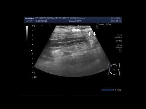
Ultrasound Video showing Fecolith in Appendix.

Показать описание
This video shows Fecolith in Appendix.
On ultrasound, a fecolith may be visualized in the appendix, where it will cast an acoustic shadow. On abdominal radiograph or CT, the radiopaque fecolith may appear laminated due to layers of calcification.
A fecolith, also known as a coprolith or stercolith, is a stony mass of compacted feces. They are most common in the descending and sigmoid colon, but may also form in the small bowel or appendix.
Fecoliths differ in their presentation based on size and location and may first be detected with the occurrence of severe complications. Initial symptoms mostly include pain due to the partial or complete obstruction of a lumen; constipation is common but may be disguised by the presence of overflow diarrhea. Rarely, more severe symptoms result from complications; anuria, obstructed labor, and nerve compression symptoms are among those previously reported.
The factors stimulating the formation of Fecoliths are unknown but may include ingested foreign bodies or gallstones. Rarely, a fecolith may become very large and form a fecaloma with a mass effect on surrounding structures.
On ultrasound, a fecolith may be visualized in the appendix, where it will cast an acoustic shadow.
Ultrasound can identify an enlarged appendix or an abscess. Nevertheless, during appendicitis, an enlarged inflamed appendix or abscess can be seen in only 50% of patients. Therefore, not seeing the appendix during an ultrasound does not exclude appendicitis.
Ultrasound should be the first imaging modality for diagnosing acute appendicitis. Primary Ultrasound for Acute appendicitis. A diagnosis will decrease ionizing radiation and cost. The sensitivity of Ultrasound to diagnose acute appendicitis is lower than of CT/MRI. Non-visualization of the appendix should lead to a clinical reassessment.
Appendicitis: Sonographic criteria for a diagnosis of appendicitis include a tubular structure that is more than 6 mm in diameter, noncompressible, and lacks peristalsis.
In the absence of a distinctly visualized appendix and secondary inflammatory changes, the incidence of acute appendicitis is low. Non visualization of the appendix even when a small amount of fat is present in the right lower quadrant may safely exclude acute appendicitis if no secondary CT findings are present.
Appendicitis symptoms may last between 36 to 72 hours before the appendix ruptures. Appendicitis symptoms develop quickly from the onset of the condition. Early symptoms include pain near the belly button, loss of appetite, nausea, and vomiting, and a low fever.
 0:04:43
Ultrasound Video showing Fecolith in Appendix.
 0:03:07
Ultrasound Video showing Inflamed Appendix with a fecolith in it.
 0:07:39
Ultrasound Video showing a case of Appendicitis with fecolith and lymph nodes.
 0:02:14
Ultrasound Video showing an inflamed appendix with fecolith.
 0:01:52
Ultrasound Video showing Inflamed Appendix with fecolith seen in its interior.
 0:04:51
Ultrasound Video showing an inflamed appendix with a fecalith in it.
 0:00:20
ultrasound video showing inflamed appendix with fecolith
 0:07:13
The Scanning of the inflamed Appendix with fecolith in its interior.
 0:02:16
Ultrasound Video showing an inflamed Appendix.
 0:03:59
Ultrasound Video showing the Inflamed Appendix and Cholelithiasis.
 0:05:22
Scanning and Focusing of Inflamed Appendix with Fecolith seen in its lumen.
 0:03:38
Ultrasound Video showing the focusing and scanning of acute appendicitis.
 0:04:31
The diagnosis of Acute Appendicitis with fecolith by Ultrasound.
 0:03:01
Ultrasound Video showing a case of Acute Appendicitis.
 0:01:59
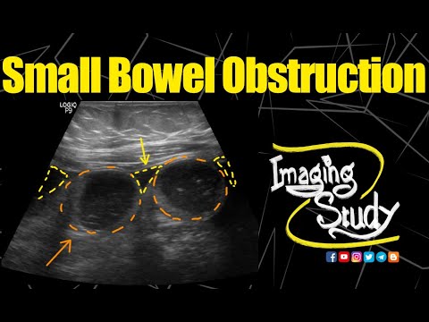
Small Bowel Obstruction || Ultrasound || Case 152
 0:04:13
Ultrasound Video showing the technique to localize the Inflamed Appendix.
 0:04:14
Ultrasound Video showing Inflamed appendix with faecolith in its interior.
 0:03:09
Ultrasound Video showing focusing of acute appendicitis.
 0:08:22
Ultrasound Video showing scanning of Inflamed Appendix.
 0:04:51
Ultrasound Video showing focusing of Acute Appendicitis.
 0:00:22
Ultrasound video showing a case of sub-hepatic acute appendicitis
 0:01:17
Ultrasound Video showing an inflamed Appendix
 0:05:28
Ultrasound Video showing localization of inflamed Appendix in a male child aged about 10 years.
 0:03:04
Ultrasound Video showing Focusing of the inflamed Appendix.
Комментарии