filmov
tv
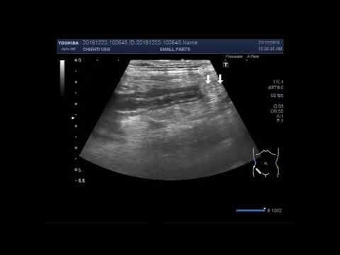
Ultrasound Video showing localization of inflamed Appendix in a male child aged about 10 years.

Показать описание
This video shows localization of inflamed Appendix. A small amount of free fluid is also seen in the lower abdomen in this case.
This is a case of pain in Rt. iliac fossa in a male patient of about 10 years age. When acute appendicitis is suspected , scan the patient lying supine. I selected a high power ( from 07 to 11 MHz ) transducer. You may put a pillow under the both knees to have the abdomen of the patient relaxed. Please start the longitudinal scan in the Rt. lower abdomen. You will start by applying the gentle pressure, then apply firmer pressure to displace the bowel to avoid bowl gases. As the bowl loops are inflamed, they are fixed without peristaltic movements. Most often tenderness will help you to localize the appendix. The appendix is seen as on cross-sectional scan as concentric circle. You can see that inner lumen is hypoechogenic with hyperechogenic edema surrounding it. Now if you scan in long axix, appendix will appear in tubular form with the same pattern.
It is not easy to localize the appendix but once you practice it, it looks much easy. Practice makes a man perfect you know
 0:05:28
 0:04:13
 0:09:47
 0:08:28
 0:06:17
 0:08:22
 0:05:04
 0:00:30
 0:03:38
 0:00:31
 0:04:20
 0:03:01
 0:03:59
 0:04:52
 0:04:51
 0:05:56
 0:06:13
 0:05:31
 0:03:03
 0:02:52
 0:05:38
 0:06:55
 0:00:17
 0:09:01