filmov
tv
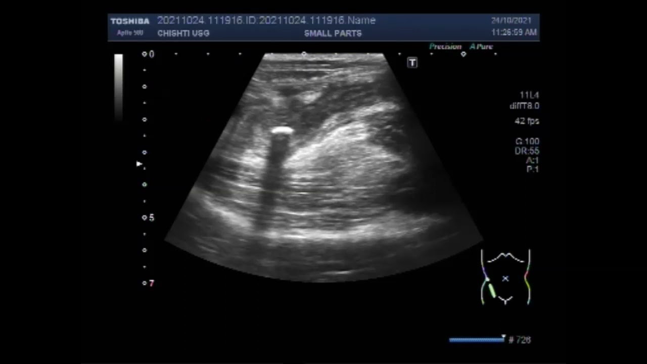
Scanning and Focusing of Inflamed Appendix with Fecolith seen in its lumen.
Показать описание
This video shows Scanning and Focusing of Inflamed Appendix with Fecolith seen in its lumen.
Appendicitis is a painful swelling of the appendix. The appendix is a small, thin pouch about 5 to 10cm (2 to 4 inches) long. It's connected to the large intestine, where poo forms. Nobody knows exactly what the appendix does, but removing it is not harmful.
Not all people will have the same symptoms, but you must see a doctor as quickly as possible. According to Johns Hopkins Medicine, the appendix can rupture as quickly as 48 to 72 hours after the onset of symptoms.
Consensus dictates that the main etiology of appendicitis is obstruction secondary to fecalith formation within the lumen of the appendix in adults. Other uncommon causes may include parasites, undigested plant or fruit residues, trauma, and foreign bodies.
Fecalith: A hard stony mass of feces in the intestinal tract. A fecalith can obstruct the appendix, leading to appendicitis. Fecaliths can also obstruct diverticuli. Also known as coprolith and stercolith. It is also called appendicolith when it occurs in the appendix.
An appendicolith is a calcified deposit within the appendix. They are present in a large number of children with acute appendicitis and may be an incidental finding on an abdominal radiograph or CT. Incidence may be increased among patients with a retrocecal appendix.
By using the high-frequency probe i.e. High power linear probe, You can see a tubular structure in the Rt. Iliac fossa surrounded by a hypoechoic rim. This is the transverse view. Now you can see a small amount of fluid is surrounding this tubular structure. Now by sweeping the probe I can see a longitudinal view of this tubular structure. Bowl walls are thin. Both ends of this tubular structure are blunt. By applying some pressure with the probe you can see this structure is not compressible. It does not collapse. A significant mucus and debris are seen in its interior but no Fecolith is seen in this case. You may find faecolith in some other cases. This area is extremely tender with the patient experiencing intense pain.
Appendicitis happens when the inside of your appendix is blocked. Appendicitis may be caused by various infections such as viruses, bacteria, or parasites, in your digestive tract. Or it may happen when the tube that joins your large intestine and appendix is blocked or trapped by stool.
Most of the patients with appendicolith are asymptomatic. However, an appendicolith may be associated with complicated appendicitis with serious outcomes.
A blockage in the lining of the appendix that results in infection is the likely cause of appendicitis. The bacteria multiply rapidly, causing the appendix to become inflamed, swollen, and filled with pus. If not treated promptly, the appendix can rupture.
It is generally accepted that the main etiology of appendicitis is obstruction due to fecalith in adults and lymphoid hyperplasia in children. It is also accepted that perforated/gangrenous appendicitis is associated with an obstructed appendix secondary to the presence of a fecalith.
The most useful sign of acute appendicitis on ultrasonography is an outer appendiceal diameter of 6 mm or greater on cross-section. Depending on the technique used, the diagnostic accuracy of CT in acute appendicitis ranges from 93 to 98 percent.
Sometimes stool can get stuck in the appendix, which is shaped like a tube with one closed end. Like a balloon that's been tied off, there's no way for what's trapped inside to escape. The pressure builds as the appendix continues producing its normal secretions.
Infection is one of the most common causes of appendicitis. A viral or bacterial infection causes the appendix to swell and fill with pus. The inflammation blocks blood flow to the appendix, which then starts to die. At this point, the appendix can develop holes or tears or may even burst if it is not treated.
Appendicitis can be acute or chronic. In acute cases of appendicitis, the symptoms tend to be severe and develop suddenly. In chronic cases, the symptoms may be milder and may come and go over several weeks, months, or even years.
Appendicitis symptoms may last between 36 to 72 hours before the appendix ruptures. Appendicitis symptoms develop quickly from the onset of the condition. Early symptoms include pain near the belly button, loss of appetite, nausea, and vomiting, and a low fever.
The most telltale symptom of appendicitis is a sudden, sharp pain that starts on the right side of your lower abdomen. It may also start near your belly button and then move lower to your right.
Symptoms of Appendicitis
Pain in your lower right belly or pain near your navel that moves lower. This is usually the first sign.
Loss of appetite.
Nausea and vomiting soon after belly pain begin.
Swollen belly.
Fever of 99-102 degrees.
Can't pass gas.
 0:05:22
Scanning and Focusing of Inflamed Appendix with Fecolith seen in its lumen.
 0:04:56
Ultrasound Video showing Focusing of inflamed Appendix.
 0:08:22
Ultrasound Video showing scanning of Inflamed Appendix.
 0:07:13
The Scanning of the inflamed Appendix with fecolith in its interior.
 0:06:59
Ultrasound Tutorial: Appendix/Appendicitis | Radiology Nation
 0:45:22
Inflammation and Infection Imaging webinar
 0:03:05
Ultrasound Video showing focusing of inflamed appendix.
 0:02:35
Acute appendicitis on CT - radiology video tutorial
 0:03:04
Ultrasound Video showing Focusing of the inflamed Appendix.
 0:03:10
Ultrasound Video showing the Focusing and localizing of inflamed Appendix.
 0:20:52
Jarred Younger, PhD | How Brain Inflammation Causes ME/CFS
 0:02:41
Crohn's Disease - Active Inflammation Overview
 0:00:11
When Appendix becomes Infected and Complicated #ultrasound #appendicitis #intestine
 0:32:34
Lecture 9 Infection and inflammation imaging
 0:04:02
Inflamed Appendix and a stone in the ureter.
 0:00:42
What is inflammation? and why does it happen?
 0:17:02
The Age of Human Language & Scanning for Inflammation in Dementia | Scilights
 0:00:25
Unraveling the Difference Between Inflammation and Cancer Cells Decoding PET Scan Results#health
 0:49:52
Webinar 11/2020: Imaging skin inflammation by means of Raster-Scanning Optoacoustic Mesoscopy (RSOM)
 0:03:40
Swollen Occipital Lymph Node: What's Behind It?
 0:02:34
Diagnosing and Treating Pituitary Tumors - California Center for Pituitary Disorders at UCSF
 0:12:07
Inflamed Appendix with Sealed perforation and enlarge Lymph nodes.
 0:24:30
Using Molecular Imaging to Study Inflammation
 0:04:50
What happens to your brain during a migraine - Marianne Schwarz
Комментарии