filmov
tv
Wrist MRI Anatomy | Radiology anatomy part 1 prep | How to interpret a wrist MRI

Показать описание
*High yield radiology physics past paper questions with video answers*
Perfect for testing yourself prior to your radiology physics exam 👇
➡️ MRI QUESTION BANK: COMING SOON 🕰️
=========================
*I have also created two RADIOPAEDIA LEARNING PATHWAYS*
WHAT’S INCLUDED?
✅This YouTube series Ad free
✅Constantly updated Radiopaedia articles
✅Summary slides
✅Key take home bullet points throughout
✅Multiple review quizzes
✅Short answer review questions
✅Official Radiopaedia course completion certificate
=========================
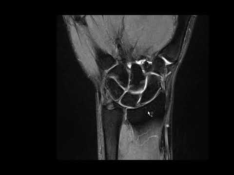
Our MSK imaging tutorials continue. Today we review wrist MRI anatomy.
We’ll cover the bones of the forearm, carpal bones, dorsal wrist tendons, flexor tendons as well as intrinsic and extrinsic wrist ligaments. We also review the triangularfibrocartilage complex (including the triangular fibrocartilage disk proper, the ulnomeniscal homologue and the ligaments that surround the TFCC.
Easily identify these structures: radius, ulna, scaphoid, lunate, triquetrum, hamate, pisiform, capitate, trapezoid, trapezium, flexor digitorum profundus and superficialis tendons, the six dorsal extensor tendon compartments and their contents, the intrinsic wrist tendons (scapholunate, lunotriquetral) and extrinsic wrist tendons (radiocarpal tendons, ulnocarpal tendons).
Chapters:
0:00 Intro
0:57 Overview of wrist bones
4:22 Carpal bone anatomy
5:52 Tendons(flexor and extensor) on axial MRI
11:44 Median and ulnar nerve
12:08 Overview of wrist ligaments
13:01 Intrinsic ligaments (CT and MRI)
17:10 Volar extrinsic ligaments
21:00 Dorsal extrinsic ligaments
23:24 Triangular fibrocartilage complex (TFCC)
26:47 Summary
=========================
*Not sure if the question banks are for you?*
If you're here, you're likely studying for a radiology physics exam. I've spent the last few months collating past papers from multiple different countries selecting the most commonly asked questions. You'll be surprised how often questions repeat themselves!
The types of questions asked in FRCR, RANZCR AIT, ARRT, FC Rad Diag (SA), ABR qualifying Core Physics and MICR part 1 are surprisingly similar and the key concepts remain the same throughout. I've taken the most high-yield questions and answered them in video format so that I can take you through why certain answers are correct and others are not.
Happy studying,
Michael
#radiology #radres #FOAMrad #FOAMed
Perfect for testing yourself prior to your radiology physics exam 👇
➡️ MRI QUESTION BANK: COMING SOON 🕰️
=========================
*I have also created two RADIOPAEDIA LEARNING PATHWAYS*
WHAT’S INCLUDED?
✅This YouTube series Ad free
✅Constantly updated Radiopaedia articles
✅Summary slides
✅Key take home bullet points throughout
✅Multiple review quizzes
✅Short answer review questions
✅Official Radiopaedia course completion certificate
=========================
Our MSK imaging tutorials continue. Today we review wrist MRI anatomy.
We’ll cover the bones of the forearm, carpal bones, dorsal wrist tendons, flexor tendons as well as intrinsic and extrinsic wrist ligaments. We also review the triangularfibrocartilage complex (including the triangular fibrocartilage disk proper, the ulnomeniscal homologue and the ligaments that surround the TFCC.
Easily identify these structures: radius, ulna, scaphoid, lunate, triquetrum, hamate, pisiform, capitate, trapezoid, trapezium, flexor digitorum profundus and superficialis tendons, the six dorsal extensor tendon compartments and their contents, the intrinsic wrist tendons (scapholunate, lunotriquetral) and extrinsic wrist tendons (radiocarpal tendons, ulnocarpal tendons).
Chapters:
0:00 Intro
0:57 Overview of wrist bones
4:22 Carpal bone anatomy
5:52 Tendons(flexor and extensor) on axial MRI
11:44 Median and ulnar nerve
12:08 Overview of wrist ligaments
13:01 Intrinsic ligaments (CT and MRI)
17:10 Volar extrinsic ligaments
21:00 Dorsal extrinsic ligaments
23:24 Triangular fibrocartilage complex (TFCC)
26:47 Summary
=========================
*Not sure if the question banks are for you?*
If you're here, you're likely studying for a radiology physics exam. I've spent the last few months collating past papers from multiple different countries selecting the most commonly asked questions. You'll be surprised how often questions repeat themselves!
The types of questions asked in FRCR, RANZCR AIT, ARRT, FC Rad Diag (SA), ABR qualifying Core Physics and MICR part 1 are surprisingly similar and the key concepts remain the same throughout. I've taken the most high-yield questions and answered them in video format so that I can take you through why certain answers are correct and others are not.
Happy studying,
Michael
#radiology #radres #FOAMrad #FOAMed
 0:28:16
Wrist MRI Anatomy | Radiology anatomy part 1 prep | How to interpret a wrist MRI
 0:08:08
MRI Anatomy of TFCC
 0:21:58
MRI wrist
 0:14:25
MRI WRIST TUTORIAL
 0:21:35
Elbow MRI Anatomy | Radiology anatomy part 1 prep | How to interpret an elbow MRI
 0:39:48
Wrist MRI (Approach to MSK MRI Series)
 0:36:03
Interpretation of Wrist MRI: Detailed Anatomy
 0:10:58
MRI Anatomy of Wrist by Dr Suvinay Saxena @ConceptualRadiology
 0:10:10
MRI Online: Wrist Part IX
 0:26:48
Wrist Extensor Compartments - Anatomy and Imaging Pathologies | Radiology Capsules | Episode 1
 0:05:47
How to Assess Wrist MRI
 0:07:31
MRI Online: Wrist, Part VII
 0:17:50
Wrist bones and alignment | Radiology anatomy part 1 prep | Wrist X-ray interpretation
 0:08:53
MRI Online: Wrist Part V
 0:14:45
CT MRI WRIST ANATOMY Dr/AHMED EISAWY
 0:03:31
Wrist MRI Anatomy Atlas
 0:06:01
Hand MRI Anatomy Atlas
 0:00:54
MRIs Are Insane
 0:06:10
Wrist MRI Part 1: Carpal Bones
 0:14:45
CT MRI WRIST ANATOMY
 0:02:28
Wrist MRI Masterclass
 0:12:42
MRI HAND TUTORIAL: HOW TO READ A MRI HAND EXAM?
 0:09:35
MRI Online: Wrist Part VI Evaluating Impingement Syndromes of the Wrist
 0:12:56
MRI Online: Wrist MRI Protocol Wrist, Part II
Комментарии