filmov
tv
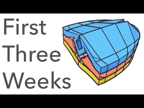
Embryology Animated - the First Three Weeks

Показать описание
Embryology animation in 3D is essential, because embryology is a difficult topic to get your head around. I've tried to make it as easy as possible. In this video I try to answer questions like: What is embryology? What happens in the first three weeks? What is gastrulation and neurulation?
#3D_Embryology
#######
Video Script
#######
In this video, we take a simplified tour through the first three weeks of embryology. Each day finishes with some etymology, because understanding of the language behind embryology is crucial to comprehending the topic well.
Week 1
Day 1
The egg is invaded by a sperm, together forming the zygote. It contains male and female pronuclei. It’s cytoplasm is surrounded by a tough glycoprotein shell.
Zygote means ‘two things joined in a close relationship’, because sperm and egg have joined to become one cell.
Day 2
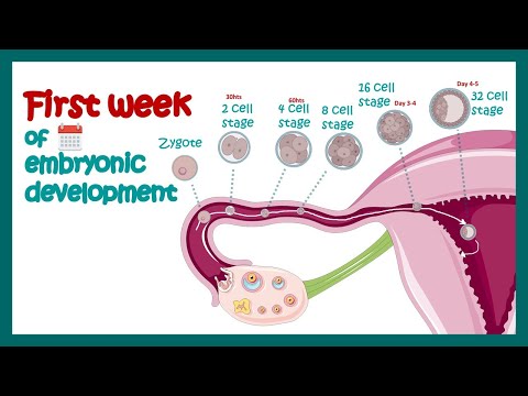
First mitotic division of the zygote occurs around 30 hours post-conception, producing two blastomeres.
Blastomere means sprout-segment, because one blastomere is one segment of the developing human-sprout.
Day 3
Division continues, and once 16 blastomeres are present they are together known as the morula, which means ‘mulberry’, because this bunch of cells visually resembles a mulberry.
Day 4
Fluid enters the morula, forming an internal cavity. The morula is now known as blastocyst. From now on, we will stop drawing individual cells, and will just show the overall shapes of structures.
Blastocyst ’sprout, again, and cyst just means ‘small fluid-filled sac’, because the human-sprout now has fluid within it.
Day 5
By day 5, the blastocyst has shed its outer shell. Cells have separated into two distinct areas: the trophoblast, which develops into the placenta and will feed the embryo, and the inner cell mass, which develops into the embryo proper.
Trophoblast comes from trephein, meaning ‘to feed’, because the trophoblast becomes the placenta which feeds the developing embryo.
Day 6
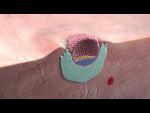
Trophoblast cells break down barriers in the uterine epithelium, allowing entry of the blastocyst.
Day 7
Meanwhile, the trophoblast begins to transform into two separate parts: cytotrophoblast on the inside, syncytiotrophoblast on the outside. The latter is one cell with many nuclei.
Syncytiotrophoblast is appropriate because it is not many separate cells, it is one ‘together-cell’.
Week 2
To simplify things, and match the imagery in most textbooks, we'll remove the endometrium now, and rotate our view like so.
Day 8
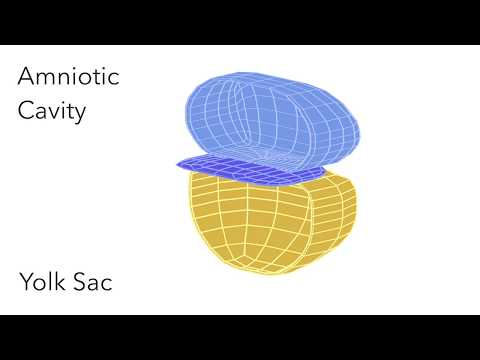
The inner cell mass differentiates into hypoblast and epiblast. A space develops in the epiblast - the amniotic cavity.
Hypoblast: meaning lower ‘sprout’
Epiblast: meaning upper sprout’
Amniotic: comes from amnion, Latin for “membrane around a foetus”.
Day 9
Meanwhile, cells of the hypoblast have migrated to replace the blastocyst cavity with the primitive yolk sac. Hypoblast and epiblast together are here known as the bilaminar disc.
Bilaminar: means 'two-layered'
Day 10
By day 10, the blastocyst is now firmly implanted in the endometrium. The syncytiotrophoblast breaks into maternal capillaries known as sinusoids, effectively co-opting their blood for the developing embryo.
Day 11
On the inner surface of the cytotrophoblast, tissue known as extra-embryonic mesoderm has developed. This eventually develops cavities which form the extra-embryonic coelom. The result is extra-embryonic mesoderm adhering to cytotrophoblast and amniotic sac, where it is known as somatopleuric, and to the yolk sac, where it is known as splanchnopleuric.
Splanchnopleuric basically means ‘on the side of the visceral organs’, because this mesoderm adheres to the viscera-side (the yolk sac becomes the gastrointestinal system).
Somatopleuric means on the side of the body.
Day 13
The space known as the extra-embryonic coelom has expanded, and is now known as the chorionic cavity. The extra embryonic mesoderm linking the cytotrophoblast to the embryo is known as the connecting stalk, which will eventually become the umbilical cord.
Chorionic: from chorion, meaning ‘outer membrane of the foetus’.
Day 14
Taking a closer look at the bilaminar disc now.
Cells in the epiblast begin to differentiate and burrow underneath. This looks like a burrow down the caudal end of the epiblast. It’s known as the primitive streak, and seeing that marks the beginning of gastrulation.
Gastrulation: means ‘becoming a gastrula (a trilaminar embryonic disc - more on that in week 3)
 0:11:49
Embryology Animated - the First Three Weeks
 0:03:35
Embryo Development Week by Week: IVF Time Lapse Journey
 0:04:53
Embryology | Fertilization, Cleavage, Blastulation | First week of embryonic development | Zygote
 0:00:35
Embryo Development _Become a baby 🍼
 0:08:52
Educational Content ,From Fertilization To Childbirth | 3d medical animation | by Dandelion Team
 0:04:21
Fetal Development 3D Animation - Infuse Medical
 0:04:17
Pregnancy: A Month-By-Month Guide | 3D Animation
 0:02:33
Do you know how is the Heart Formation In Embryo? heart formation embryology animation
 0:00:48
Implantation of the blastocyst
 0:03:55
Embryo Folding: Amniotic Cavity & Yolk Sac - Animated Embryology
 0:17:37
Embryology | Fertilization, Cleavage, Blastulation
 0:01:21
How Fertilization happens | 3D Animation
 0:07:36
general embryology anatomy | first week of development embryology | Johari MBBS
 0:19:35
Pregnancy - How a Wonder is Born! (Animation)
 0:03:24
Neurulation - Animated Embryology
 0:00:57
#biology #zoology || Development of chick embryo
 0:07:11
Implantation of the blastocyst | Week 2 of embryonic development | Developmental biology
 0:00:29
5 days of embryo development in 30 seconds! 😍 #ivf #biology #wantababy
 0:00:07
Baby Size in Weeks 🥰 First Trimester Embryo and Fetal Development #pregnancy #fetus #embryo
 0:00:18
What is primitive streak?
 0:00:15
1st week of life. Implantation of a Fertilized Egg. #shortvideo #baby #pregnant August 27, 2024
 0:00:16
What Happens to the Embryonic Tail as We Develop? 🤔
 0:00:14
Embryo development process in IVF #embryology #embryodevelopment #embryologist #ivf #ivfprocess
 0:04:34
DPES EarlyEmbryonicFacialDevelopment
Комментарии