filmov
tv
Normal Skin Histology - Explained by a Dermatopathologist

Показать описание
Topics discussed:
Epidermis:
Layers of epidermis: 0:10
Melanocytes vs Keratinocytes: 5:16
Langerhans cells: 10:10 & 33:30 & 57:30
Dermis:
Papillary and reticular dermis: 11:50
Three types of white empty spaces on a slide: vessels, glands/ducts/cysts, or artifact: 15:25
Blood vessels & nerves: 18:24 & 48:50 & 58:59
Arrector pili & other dermal smooth muscle: 20:00
Adnexal:
Sebaceous gland: 21:10
Hair follicle 23:14
Eccrine sweat glands and ducts 24:45 & 50:00
Gland/duct vs blood vessel 27:20 & 48:50
Three types of pink bundles: smooth muscle, nerve, dense connective tissue: 27:50
Acral skin (palm sole) with contact dermatitis 29:37
Parakeratosis 30:00
Perivascular lymphocytes 30:40
Eosinophils vs neutrophils 31:20
Spongiosis with desmosome keratinocyte spines 32:10
Spongiotic vesicles with Langerhans cells 33:30
Normal acral skin (palm & sole) with stratum lucidum 34:20
Normal glomus body/apparatus (canal of Sucquet-Hoyer) 35:40
Nerve 36:46 & 51:50
Adipose tissue (white fat cells) in subcutis with Lochkern 37:55
Normal scalp skin with large anagen hair follicles: 39:30
Hair follicle anatomy (bulb/matrix, inner root sheath, outer root sheath, hair shaft, isthmus, infundibulum): 40:55 (labeled images):
Pacinian corpuscle 50:40
Meissner corpuscle 1:02:28
Dense regular connective tissue (Fascia/Tendon/Ligament) vs Smooth Muscle 53:00
Basic Normal Skin Immunohistochemistry:
-cytokeratin in epidermis: 55:33
-S100 in melanocytes and Langerhans cells and adipocytes: 57:30
-Desmin in smooth muscle (arrector pili and blood vessels): 58:59
-CD31 in endothelial cells of blood vessels: 59:33
-SOX-10 in melanocytes: 1:00:40
Digit/Finger/Toe histology (amputation for subungual acral melanoma) 1:04:10 & 1:08:30
-bone 1:05:40
-glomus body 1:05:15
-tendon/ligament 1:06:10
-artery 1:06:58
-fingernail/toenail 1:08:54
-acrosyringium 1:10:45
Solar elastosis (what wrinkles look like microscopically!) 1:11:50
Other videos you might like:
The basic normal structures of the skin discussed and described by a dermatopathologist. This material is intended for use by medical students, junior pathology or dermatology residents, or for anyone else studying normal human histology. Special thanks to two of my medical students at UAMS for helping make this video possible. Miki Lindsey convinced me that I really needed to sit down and record this video. Akash Patel took time to edit the video and make it ready for YouTube. My sincere thanks to both of them for helping me overcome procrastination.
Correction - I made a mistake in the video. I said that sebaceous gland secretions are turned into smelly substances by bacteria and that this makes body odor. That is incorrect. That is actually true of APOCRINE gland secretions not sebaceous secretions.
Also, in the past I used "keratinocyte" and "squamous cell" interchangeably (this is because in dermatopathology, we see and talk about squamous cell carcinomas all the time, and those tumors are composed of keratinocytes). But technically, in normal skin histology, "squamous cell" refers only to the flattened keratinocytes in the superficial epidermis. Thankfully, a histology PhD colleague pointed this out to me and corrected my lazy nomenclature!
This video is geared towards medical students, pathology or dermatology residents, or practicing pathologists or dermatologists. Of course, this video is for educational purposes only and is not formal medical advice or consultation.
Presented by Jerad M. Gardner, MD. Please subscribe to my channel to be notified of new pathology teaching videos.
Follow me on:
Snapchat: JMGardnerMD
Twitter: @JMGardnerMD
Instagram: @JMGardnerMD
 1:14:19
Normal Skin Histology - Explained by a Dermatopathologist
 0:10:32
Skin: Histology
 0:04:03
Histology of the Skin
 0:16:23
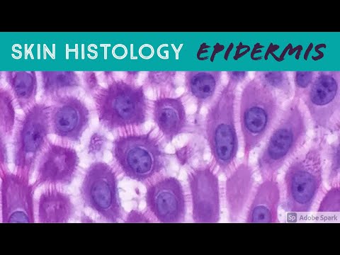
Skin Histology: Epidermis Layers (stratum basale, spinosum, granulosum, lucidum & corneum)
 0:37:43
Introduction to Histology
 0:11:47
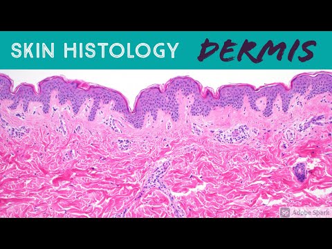
Skin Histology: DERMIS (the Epidermis can't live without it!)
 0:08:28
Epithelial Tissue Histology Explained for Beginners | Corporis
 0:24:38
Skin Histology [Integumentary System Histology Part 1 of 2]
 0:09:46
Skin Histology: Acral Skin (aka glabrous skin of palm of hand & sole of foot)
 0:09:40
The Integumentary System, Part 1 - Skin Deep: Crash Course Anatomy & Physiology #6
 0:13:40
Identifying Epithelium | Review and Practice Questions
 0:19:59
Histology of the Skin part 1
 0:07:58
Kidneys: Histology
 0:03:45
3 Min Histology - Epidermis Part 1
 0:04:00
Histology of thin skin, slide identification and explanation
 0:07:54
Connective Tissue Histology Explained for Beginners | Corporis
 0:09:36
Histology- Cancer
 0:01:42
Skin Histology: PACINIAN CORPUSCLE (MY FAVE!!!)
 1:37:54
Normal skin histology, special stains, and basic immunohistochemical stains.
 0:12:43
The Skin | The Largest Organ of Your Body | Anatomy & Histology
 0:05:25
Histology of Thick Skin/Glabrous skin
 0:43:09
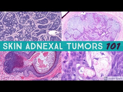
Skin Adnexal Tumors 101: A Basic Approach for General Pathologists
 0:06:40
Skin Histology: Immunohistochemistry Basics (for Dermatology Histotechnologist Dermpath Pathology)
 0:49:46
Skin. Histology
Комментарии