filmov
tv
History of X-rays

Показать описание
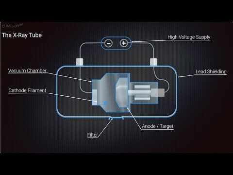
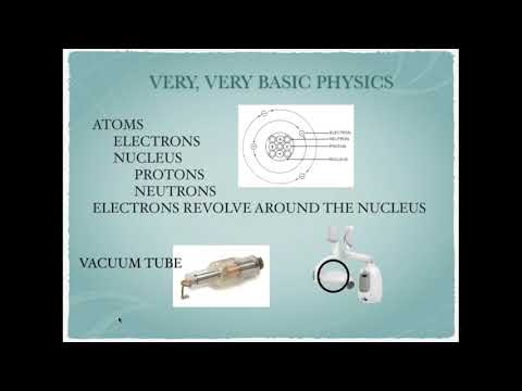
Discusses the discovery, development and basic physics of x-ray generation.
 0:24:21
History of X-rays
 0:02:23
The history of X-ray: a journey through time and medical technology
 0:04:42
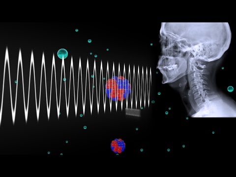
How X-rays see through your skin - Ge Wang
 0:11:12
The HORRIFIC History of X-Rays
 0:01:35
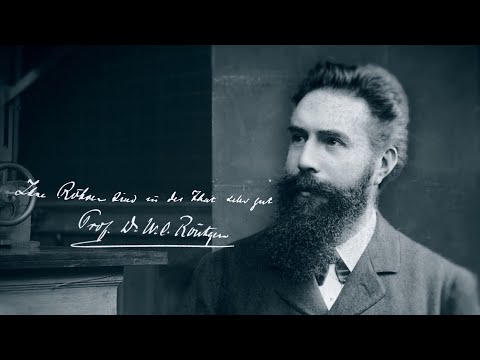
Wilhelm Conrad Röntgen and the discovery of the X-rays
 0:01:29
How Do X-rays Work?
 0:07:00
The history of Radiography
 0:04:07
How Were X Rays Discovered by Accident?
 0:09:13
SCIATICA PAIN RELIEF TREATMENT, 5 Step Non-Surgical Treatment for Sciatica | Dr. Ruminder Birk
 0:02:12
Production of X Rays animated
 0:06:01
How do X-Rays Work?
 0:09:04
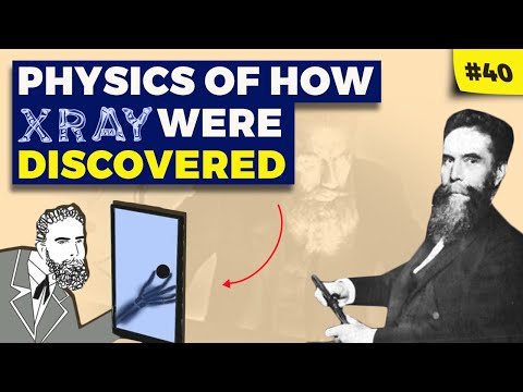
Physics of How Wilhelm Roentgen Discovered X-rays
 0:08:03
History of x rays
 0:01:50
The History of X-Rays in Medicine | Then vs. Now | GoodRx
 0:03:46
How Dangerous Are X-Rays?
 0:01:07
How do X-rays work? | #aumsum #kids #science #education #children
 0:02:11
The History of Radiology: Discovering X-Rays
 0:03:52
History and Properties of X-Rays
 0:07:29
X Ray Production Animation
 0:02:30
The Strange History of Soviet X-Ray Records
 0:05:29
Invention Of X-Ray | The Dr. Binocs Show | Best Learning Video for Kids | Preschool Learning
 0:26:18
History & Production of Dental X-rays
 0:02:23
Wilhelm Röntgen: X-rays | Heroes of Progress | Ep. 26
 0:13:54
From Rontgen to Radiology: The History of X-Rays (Mini Documentary)
Комментарии