filmov
tv
Right Atrium - Location, Anatomy & Function - Human Anatomy | Kenhub
Показать описание
Oh, are you struggling with learning anatomy? We created the ★ Ultimate Anatomy Study Guide ★ to help you kick some gluteus maximus in any topic. Completely free. Download yours today:
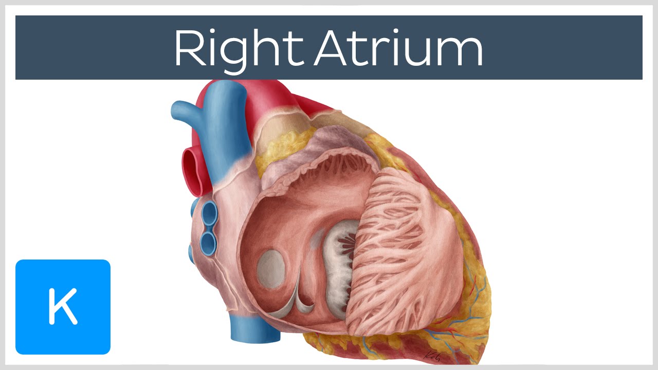
The right atrium receives deoxygenated venous flow from the superior vena cava, the inferior vena cava, the coronary sinus and the anterior and smallest cardiac veins. It passes the blood to the right ventricle of the heart through the tricuspid valve, which has three cusps or leaflets, and is also known as the right atrioventricular valve.
The main anatomical features of the right atrium include the sinus venarum, which surrounds the openings of the superior and inferior vena cavae as well as the coronary sinus, which in turn are also major structures. The atrial walls are made up of pectinate muscles which form a pouch known as the right auricle. The crista terminalis separates the cardiac and smooth muscle layers of the atrial walls, while the interatrial septum divides the atria.
0:17 Functions of the right atrium
0:52 Anatomy of the right atrium
1:32 Pectinate muscles
Want to test your knowledge on right atrium? Take this quiz:
 0:02:18
Right Atrium - Location, Anatomy & Function - Human Anatomy | Kenhub
 0:08:13
Heart Anatomy - Right Atrium - 3D Anatomy Tutorial
 0:19:32
Right atrium anatomy | External and internal features of right atrium
 0:04:18
Heart: Right atrium
 0:19:42
Right atrium anatomy | External and internal features of right atrium| [Simplified]
 0:18:19
Anatomy of the Heart - External & Internal Structures
 0:11:06
Path of Blood Flow through the Heart | Step by step through every chamber, valve, and major vessel
 0:00:05
Heart Chambers #heart #heartanatomy #anatomy #cardiology #animation #shorts
 0:34:55
Cardiovascular system physiology - Part 5
 0:00:16
Anatomy of the Heart: Size and Location ❤️
 0:00:10
Heart valves function #heartbeat# anatomy and physiology#
 0:04:54
Right Atrium Anatomy - Explained in Mixed Reality
 0:05:32
Right Atrium
 0:00:23
Heart Sounds 🔈🫀
 0:00:05
Heart anatomy and coronary artery l IVC l SVCl Aortal #heart #shorts
 0:03:35
HEART ANATOMY in 3 MINUTES| Memorize parts of the heart
 0:00:21
Heart Location & Anatomy 🫀
 0:30:14
RIGHT ATRIUM OF HEART : INTERNAL FEATURES
 0:16:57
Right Atrium of Heart (1/2) | External Features | Anatomy | EOMS
 0:21:33
Cardiovascular System 1, Heart, Structure and Function
 0:00:18
basics of heart
 0:00:16
Best Heart Location for Blood Supply 🫀
 0:03:29
Right atrium and ventricle overview (preview) - Human Anatomy | Kenhub
 0:01:01
Basic Blood Flow Through the Heart | TEAS and MCAT Review #shorts #anatomy
Комментарии