filmov
tv
Structure Of The Small Intestine - Functions Of The Small Intestine - What Are Villi

Показать описание
In this video we discuss the structure and functions of the small intestine. We also cover the villi and microvilli and the different types of cells of the small intestine.
Structure and functions of the small intestine
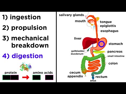
Your small intestine is an unbelievable organ that has many important roles in everyday bodily functions. When you chew food, then swallow it, it enters your esophagus, which connects with and passes the food to your stomach, which connects with and passes it to your small intestine.
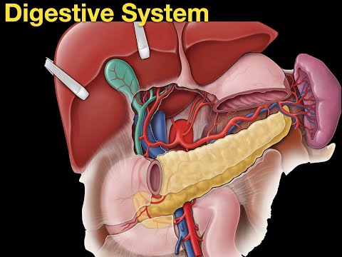
And, your small intestine is located, just below your stomach as it coils and loops filling a large portion of your abdominal cavity. The small intestine has 3 sections, with the duodenum being the first section and it wraps around the pancreas and connects to the pylorus of the stomach. The jejunum is the 2nd middle region of the small intestine and the ileum being the 3rd section. At the end of the ileum is the ileocecal valve, which controls the entry of materials into the large intestine.
The wall of the small intestine is comprised of a mucosa or mucous membrane, a submucosa , a layer of circular muscle, a layer of longitudinal muscle and a serosa. The mucosa and submucosa have internal circular folds as you can see here in this photo of the duodenum.
These folds are more numerous in the duodenum and jejunum and they increase the surface area which slows down the movement of materials from the stomach, and also provides for more nutrient digestion and absorption.
The structure of these folds is absolutely amazing as they have fingerlike projections called villi.
Each individual villus contains a rich capillary network as well as a lacteal which is a lymphatic capillary. The capillaries absorb most of the nutrients and the lacteal absorbs lipids, or fats, and lipid soluble vitamins. There are mainly 2 types of cells that line the surface of a villus.
Absorptive cells called enterocytes, and mucous producing goblet cells. The enterocyte cells have a rapid turnover, as they are shed and excreted in the feces approximately every 3 to 5 days. The enterocytes have tiny hair like projections called microvilli, also described as the brush border. The brush border contains enzymes that complete chemical digestion or breakdown of nutrients before absorption takes place.
These microvilli increase surface area even more, providing for even greater digestion and absorption. Scattered throughout the surface of a villus are also tuft cells, which are believed to secrete endorphins and enzymes that make prostaglandins, which are a type of fat that enhances the immune system.
Deep in the valleys between villi are depressions called crypts or intestinal glands. These crypts contain enteroendocrine cells that secrete hormones, intestinal stem cells, which produce daughter cells that push upwards and become enterocytes. Paneth cells are also located here, and they protect against bacterial growth.
The small intestine has 2 important functions, digestion of nutrients and absorption of nutrients. The tremendous amount of surface area of this organ makes it perfect for this.
I will cover, in depth, the process of digestion and absorption of all of the different types of nutrients in a future video.
Timestamps
0:00 The path of food to the small intestine
0:27 The 3 sections of the small intestine
0:48 The wall and folds of the small intestine
1:17 Villi in the small intestine
1:36 Enterocyte cells and microvilli in the small intestine
2:44 The functions of the small intestine
Structure and functions of the small intestine
Your small intestine is an unbelievable organ that has many important roles in everyday bodily functions. When you chew food, then swallow it, it enters your esophagus, which connects with and passes the food to your stomach, which connects with and passes it to your small intestine.
And, your small intestine is located, just below your stomach as it coils and loops filling a large portion of your abdominal cavity. The small intestine has 3 sections, with the duodenum being the first section and it wraps around the pancreas and connects to the pylorus of the stomach. The jejunum is the 2nd middle region of the small intestine and the ileum being the 3rd section. At the end of the ileum is the ileocecal valve, which controls the entry of materials into the large intestine.
The wall of the small intestine is comprised of a mucosa or mucous membrane, a submucosa , a layer of circular muscle, a layer of longitudinal muscle and a serosa. The mucosa and submucosa have internal circular folds as you can see here in this photo of the duodenum.
These folds are more numerous in the duodenum and jejunum and they increase the surface area which slows down the movement of materials from the stomach, and also provides for more nutrient digestion and absorption.
The structure of these folds is absolutely amazing as they have fingerlike projections called villi.
Each individual villus contains a rich capillary network as well as a lacteal which is a lymphatic capillary. The capillaries absorb most of the nutrients and the lacteal absorbs lipids, or fats, and lipid soluble vitamins. There are mainly 2 types of cells that line the surface of a villus.
Absorptive cells called enterocytes, and mucous producing goblet cells. The enterocyte cells have a rapid turnover, as they are shed and excreted in the feces approximately every 3 to 5 days. The enterocytes have tiny hair like projections called microvilli, also described as the brush border. The brush border contains enzymes that complete chemical digestion or breakdown of nutrients before absorption takes place.
These microvilli increase surface area even more, providing for even greater digestion and absorption. Scattered throughout the surface of a villus are also tuft cells, which are believed to secrete endorphins and enzymes that make prostaglandins, which are a type of fat that enhances the immune system.
Deep in the valleys between villi are depressions called crypts or intestinal glands. These crypts contain enteroendocrine cells that secrete hormones, intestinal stem cells, which produce daughter cells that push upwards and become enterocytes. Paneth cells are also located here, and they protect against bacterial growth.
The small intestine has 2 important functions, digestion of nutrients and absorption of nutrients. The tremendous amount of surface area of this organ makes it perfect for this.
I will cover, in depth, the process of digestion and absorption of all of the different types of nutrients in a future video.
Timestamps
0:00 The path of food to the small intestine
0:27 The 3 sections of the small intestine
0:48 The wall and folds of the small intestine
1:17 Villi in the small intestine
1:36 Enterocyte cells and microvilli in the small intestine
2:44 The functions of the small intestine
 0:06:30
Small intestine 1: Structure | Gastrointestinal system physiology | NCLEX-RN | Khan Academy
 0:19:24
Small Intestine Anatomy (Parts, Topography, Structures, Layers)
 0:02:45
The Intestinal Villi Explained || Absorption
 0:14:31
What is the Structure and Function of Small Intestine?
 0:03:11
Structure Of The Small Intestine - Functions Of The Small Intestine - What Are Villi
 0:05:59
Small Intestine | Jejunum & Ileum
 0:04:29
Small Intestine - CBSE 11
 0:01:54
small intestine | 3d medical animation
 0:09:01
Internal Organs of Human body | Parts of The Body | Internal Organs | Body Organs in English
 0:22:32
Small intestine (anatomy)
 0:08:43
Digestive System
 0:02:08
All about the small intestine
 0:18:08
Digestive System 7, Small Intestine and absorption
 0:03:48
How the Digestive System Works | 3D Animation
 0:00:07
Villus of the small intestine / Nutrient absorption
 0:18:23
Small Intestine Structure and Layers
 0:00:51
Where is the small intestine? #anatomy
 0:14:43
The Digestive System
 0:12:51
Anatomy | Histology of the Stomach & Small Intestine
 0:41:33
Digestive system
 0:00:20
Looking Inside a Real Human Stomach | #shorts #food
 0:10:04
Introduction to the Small Intestine
 0:11:42
Digestion in small intestine
 0:18:08
Small Intestine: Structure, Digestion, Absorption
Комментарии