filmov
tv
Anatomy of the Brain | Dissectible Model

Показать описание
Ninja Nerds!
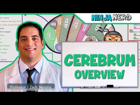
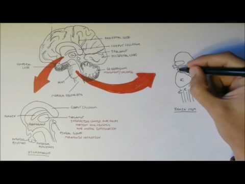
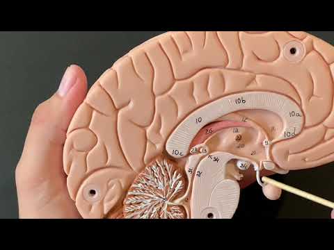
Join us in this video where Professor Zach Murphy will be discussing the anatomy of the brain through the use of a dissectible model to get a better view at smaller structures. We hope you enjoy this lecture and be sure to support us below!
Join this channel to get access to perks:
APPAREL |
We are switching merchandise suppliers.
DONATE
SOCIAL MEDIA
@NinjaNerdSci
#ninjanerd #BrainAnatomy #Neuro
 0:27:50
Anatomy of the Brain | Model
 0:32:43
Anatomy of the Brain | Dissectible Model
 0:11:58
Basic Parts of the Brain - Part 1 - 3D Anatomy Tutorial
 0:13:56
The Brain
 0:19:20
Overview of the Brain
 0:06:48
The Structure and Physiology of the Human Brain
 0:07:01
Anatomy 1, The Brain
 0:08:24
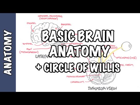
Anatomy - Brain (Circle of Willis and Stroke)
 0:00:46
'Octopus Arms Have ‘Mini-Brains’! The Incredible Intelligence of Octopuses Explained 🐙 #Shorts&...
 0:10:08
Central Nervous System: Crash Course Anatomy & Physiology #11
 0:26:34
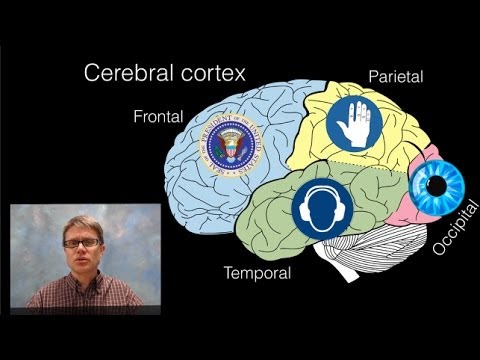
Neurology | Cerebral Cortex Anatomy & Function: Overview
 0:14:47
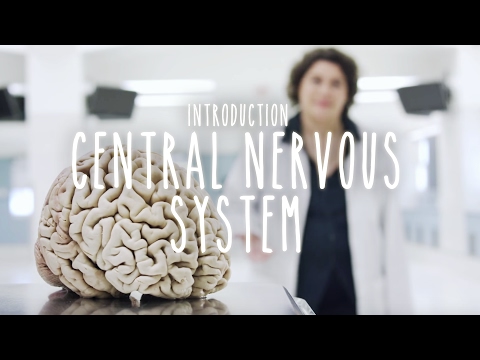
Neuroanatomy S1 E1: Intro to the Central Nervous System #neuroanatomy #science #medicine #brain
 0:10:36
Clinical Anatomy - Cerebral Cortex (lobes, injury and clinical signs)
 0:01:00
🧠 The Human Nervous System! 🧠 #brain #spinalcord #humanbody #anatomy #science #teacher #education...
 0:11:56
Brain Anatomy Overview - Lobes, Diencephalon, Brain Stem & Limbic System
 0:06:20
Divisions of the brain
 0:32:27
Introduction to Neuroanatomy - Learn the Basics - Neuroanatomy Playlist
 0:05:02
How to learn major parts of the brain quickly
 0:19:45
Lobes of the Brain: Cerebrum Anatomy and Function [Cerebral Cortex]
 0:18:43
Inside the Brain of a Cadaver | Pathways in the brain
 0:09:11
Human Anatomy, Brain Model
 0:11:54
Brain: Cerebrum and Cerebellum (+ Broca’s, Wernicke’s, and limbic overview)
 0:03:15
Ventricles of the Brain | Anatomy Model
 0:07:36
The Brain Explained for Dummies I Anatomy
Комментарии