filmov
tv
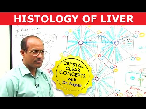
Liver Histology

Показать описание
The liver is the largest gland in the body. It is located in the upper right hand quadrant of the abdominal cavity, just inferior to the diaphragm. And the liver is subdivided into four lobes— right, left, quadrate, and caudate.
The liver is surrounded by a capsule of fibrous connective tissue called Glisson’s capsule.
Because the liver occupies a central position in metabolism, all nutrients absorbed in the alimentary canal are transported directly to the liver via the portal vein. Much of the nutritive material delivered to the liver is converted by the hepatocytes which are the chief functional cells of the liver into storage products.
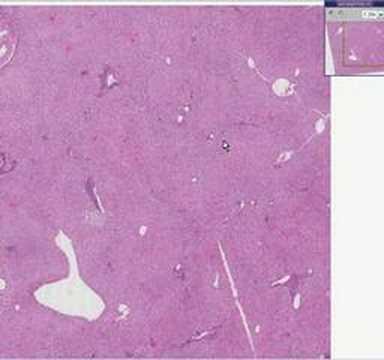
Hepatocytes are arranged in hexagon shaped lobules. Where three classical lobules are in contact with each other, the connective tissue elements are increased, and these regions are known as portal areas or portal triads. And the portal triad consists of a bile ductule, portal venule, and hepatic artery.
After identifying the lobule, it can be easier to locate portal triads in an image since they’re typically located at the corners of the lobules.
The longitudinal axis of each classical lobule is occupied by the central vein. Hepatocytes radiate, similar to spokes of a wheel, from the central vein, forming anastomosing, fenestrated plates of liver cells, separated from each other by large vascular spaces known as hepatic sinusoids.
The sinusoids carry blood from the hepatic arteriole and portal venule to the central vein, while the bile canaliculi or capillaries carry the bile produced by hepatocytes in the opposite direction in order to drain into the bile ductules.
The hepatocyte’s cytoplasm is very eosinophilic, or pink because they contain a lot of mitochondria.
Many of the cells in this image also have fine brown granules within the hepatocytes called lipofuscin.
These granules are considered a sign of “wear and tear” and it’s normal to see the amount of lipofuscin gradually increase with age.
The hepatocyte plates have a supportive tissue or stroma of reticulin fibers.
These fibers are difficult to see with an Hematoxylin and eosin stain, but a reticulin-specific stain can be used in order to visualize the fibers and hepatocyte plates better.
The liver is surrounded by a capsule of fibrous connective tissue called Glisson’s capsule.
Because the liver occupies a central position in metabolism, all nutrients absorbed in the alimentary canal are transported directly to the liver via the portal vein. Much of the nutritive material delivered to the liver is converted by the hepatocytes which are the chief functional cells of the liver into storage products.
Hepatocytes are arranged in hexagon shaped lobules. Where three classical lobules are in contact with each other, the connective tissue elements are increased, and these regions are known as portal areas or portal triads. And the portal triad consists of a bile ductule, portal venule, and hepatic artery.
After identifying the lobule, it can be easier to locate portal triads in an image since they’re typically located at the corners of the lobules.
The longitudinal axis of each classical lobule is occupied by the central vein. Hepatocytes radiate, similar to spokes of a wheel, from the central vein, forming anastomosing, fenestrated plates of liver cells, separated from each other by large vascular spaces known as hepatic sinusoids.
The sinusoids carry blood from the hepatic arteriole and portal venule to the central vein, while the bile canaliculi or capillaries carry the bile produced by hepatocytes in the opposite direction in order to drain into the bile ductules.
The hepatocyte’s cytoplasm is very eosinophilic, or pink because they contain a lot of mitochondria.
Many of the cells in this image also have fine brown granules within the hepatocytes called lipofuscin.
These granules are considered a sign of “wear and tear” and it’s normal to see the amount of lipofuscin gradually increase with age.
The hepatocyte plates have a supportive tissue or stroma of reticulin fibers.
These fibers are difficult to see with an Hematoxylin and eosin stain, but a reticulin-specific stain can be used in order to visualize the fibers and hepatocyte plates better.
 0:04:52
Liver: Histology
 0:56:05
Gastrointestinal | Liver Histology
 0:10:04
Learn Liver Histology: The Basics You Need to Know
 0:06:43
Shotgun Histology Liver
 1:35:26
Hepatocytes and Portal Vein | Liver Histology | Dr Najeeb
 0:05:51
Histology of the Liver
 0:03:22
S3DMediMagic for Histology- Histology of Liver
 0:10:55
Liver Lobules (Portal Triad)
 0:56:20
LECTURE ON -ANATOMY STOMACH BY- DR. HRISAB DEB | MBBS | TRIPURA SANTINEKITAN MEDICAL COLLEGE
 0:03:24
Liver: tissues and cells (preview) - Human Histology | Kenhub
 0:31:23
Histology of the liver
 0:06:36
Liver Histology -- Normal
 0:10:27
Histology of Liver
 0:00:58
Liver Histology #anatomy #physiology #anatomyandphysiology #premed #study #biologystudent #liver
 0:02:29
Liver Histology
 1:22:41
#LIVERPATH Liver biopsies 101: major patterns of disease
 0:29:52
Liver, Pancreas, Gallbladder Histology [GI Histology 4 of 4]
 1:01:48
Learning to love the liver logically - Part 1 - Dr. Furth (UPenn) #LIVERPATH
 0:04:58
Histopathology Liver--Normal
 0:20:59
Histology of Liver, Gall bladder & Pancreas
 1:04:07
Liver pathology: let's start with the basics - Dr. Saxena (Indiana) #LIVERPATH
 0:04:34
Hepatic Steatosis - Histopathology (+ Normal Liver Histology)
 0:50:24
5a-Histology of Liver part1-GIT
 0:38:23
Liver Histology
Комментарии