filmov
tv
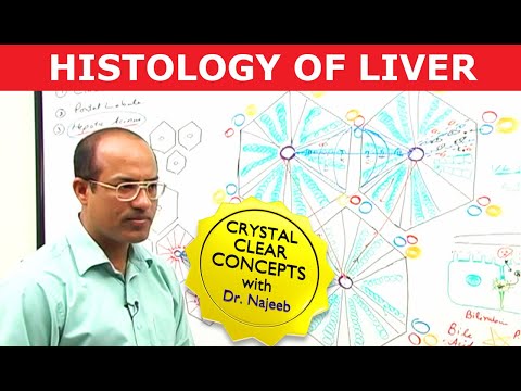
Histology of Liver

Показать описание
Sheets of connective tissue divide the liver into thousands of small units called lobules. A lobule is roughly hexagonal in shape, with portal triads at the vertices and a central vein in the middle. The lobule is the structural unit of the liver and rather easy to observe. In contrast, the hepatic acinus is more difficult to visualize but represents a unit that is of more relevance to hepatic function because it is oriented around the afferent vascular system.
Join us now for Personalized Medical Education
Download medCampus app
#medCampus #NEETPG #AIIMSPG #USMLE
Join us now for Personalized Medical Education
Download medCampus app
#medCampus #NEETPG #AIIMSPG #USMLE
 0:04:52
Liver: Histology
 0:56:05
Gastrointestinal | Liver Histology
 0:05:51
Histology of the Liver
 0:10:04
Learn Liver Histology: The Basics You Need to Know
 0:03:22
S3DMediMagic for Histology- Histology of Liver
 0:10:27
Histology of Liver
 0:10:55
Liver Lobules (Portal Triad)
 0:06:43
Shotgun Histology Liver
 1:35:26
Hepatocytes and Portal Vein | Liver Histology | Dr Najeeb
 0:31:23
Histology of the liver
 0:02:29
Liver Histology
 0:25:30
Histology of Liver
 0:20:59
Histology of Liver, Gall bladder & Pancreas
 0:03:24
Liver: tissues and cells (preview) - Human Histology | Kenhub
 0:29:52
Liver, Pancreas, Gallbladder Histology [GI Histology 4 of 4]
 0:00:58
Liver Histology #anatomy #physiology #anatomyandphysiology #premed #study #biologystudent #liver
 0:50:24
5a-Histology of Liver part1-GIT
 0:06:36
Liver Histology -- Normal
 0:07:13
Histology of liver | Microanatomy of liver 3D
 0:38:23
Liver Histology
 0:06:12
Pancreas: Histology
 0:06:05
Histology Of Liver || Histology || Chill Medicos
 0:00:36
Liver Histology Model
 0:15:02
HISTOLOGY OF LIVER 1_ Anatomy Book Club
Комментарии