filmov
tv
Cervical Spine Conditions, TUMORS - Everything You Need To Know - Dr. Nabil Ebraheim
Показать описание
Dr. Ebraheim’s educational animated video describes miscellaneous tumors of the cervical spine.
Hemangioma:
•Hemangiomas are benign hamartomatous vascular lesions in the spine.
•Most hemangiomas are isolated lesions that affect a single vertebra.
•On x-ray, hemangiomas appear as abnormal thickened trabeculae with vertical striation (honeycomb appearance).
•Most are asymptomatic and present as incidental finding and do not require intervention.
•Preoperative embolization if resection is needed.
Osteoid osteoma and osteoblastoma:
•Osteoid osteoma and osteoblastoma are histologically similar to each other, however the osteoblastoma tends to grow larger than 2 cm.
•Osteoblastoma may sometimes displace characteristics that may be confused with osteosarcoma.
•Osteoblastoma – you will do marginal excision.
•Osteoid osteoma is almost impossible to visualize onx-ray due to its small size.
•The patient usually presents painful scoliosis or torticollis (pain is relieved with aspirin).
Aneurysmal bone cyst:
•Fluid level
oPain could be present
oTreatment is excision
•Expansile osteolytic lesion with a thin wall containing blood-filled cystic cavities that usually effect the posterior element of the spine.
Eosinophilic Granuloma
•Caused by histiocytosis X
•Usually seen in children under the age of 10 years old.
•It is a lytic lesion of the vertebral body that will show on x-ray as a dense ring of collapsed cortical bone sandwiched between intact vertebral discs (vertebral plana).
•It is rarely symptomatic and self-limiting.
•Conservative treatment with orthosis.
•Large tumor may compress the spinal cord and is usually treated by low dose radiation therapy.
Giant Cell Tumor
•It usually occurs in the sacral region of the spine and less commonly affects the cervical spine.
•It may be difficult to distinguish giant cell tumors from other tumors of the spine on x-rays.
•This tumor is usually found in the anterior column of the spine.
•On x-ray it will appear as a radiolucent expansile lesion with a cortical shell and a bony septa.
•Recurrence is common secondary to inadequate resection.
•10% incidence of malignant transformation due to irradiation.
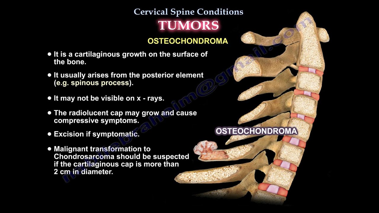
Osteochondroma
•It is a cartilaginous growth on the surface of the bone.
•It usually arises from the posterior element (eg spinous processes).
•It may not be visible on x-rays.
•The radiolucent cap may grow and cause compressive symptoms.
•Excision if symptomatic.
•Malignant transformation to chondrosarcoma should be suspected if the cartilaginous cap is more than 2 cm in diameter.
Paget’s disease
•Paget’s disease is a chronic disorder that can result in enlarged bones.
•Excessive breakdown and formation of bone tissue which ends by sclerosis giving the ivory appearance on x-ray.
•Less likely to develop in the cervical spine and more common in the sacral and lumbar regions.
•Differential diagnosis for prostatic metastasis.
Osteosarcoma
•Osteosarcoma is rare in the spine.
•Malignant tumor is found to affect any level of the spine, although the lumbar and sacral regions are more common.
•On plane x-ray, osteosarcoma appears as lytic and blastic lesions with destruction of the vertebra and relative sparing of the adjacent discs.
Ewing’s Sarcoma
•It affects the body of the vertebra.
•Most commonly detected in patients between 10-20 years of age.
•Appears on x-rays as a lytic lesion with osseous expansion or sclerosis.
Multiple Myeloma
•It is the most common primary malignancy of the bones and spine.
•It is due to malignancy of the plasma cells.
•Appears on x-rays as lytic lesions of the bone (punched out lesions).
•Skeletal survey is used to screen for other lesions throughout the body.
•Bone scans have low sensitivity in detecting disseminated disease.
•Treatment is usually radiotherapy. Surgery is used for decompression and/or stabilization.
Lymphoma
•Mainly Non-Hodgkin’s lymphoma.
•Bone lymphoma is commonly diagnosed between 40-60 years of age.
•It can be detected on plane x-rays in only 30-42% of patients.
•Treatment is radiotherapy and chemotherapy.
Metastasis
•The most common tumor that metastasis in the spine are:
oProstate neoplasm
oBreast neoplasm
oLung neoplasm
oRenal neoplasm
oThyroid tumor
•Differential diagnosis – infection
oInfection usually affects the end plates
oTumor usually affects the body and the pedicles.
Become a friend on facebook:
Follow me on twitter:
Donate to the University of Toledo Foundation Department of Orthopaedic Surgery Endowed Chair Fund:
Hemangioma:
•Hemangiomas are benign hamartomatous vascular lesions in the spine.
•Most hemangiomas are isolated lesions that affect a single vertebra.
•On x-ray, hemangiomas appear as abnormal thickened trabeculae with vertical striation (honeycomb appearance).
•Most are asymptomatic and present as incidental finding and do not require intervention.
•Preoperative embolization if resection is needed.
Osteoid osteoma and osteoblastoma:
•Osteoid osteoma and osteoblastoma are histologically similar to each other, however the osteoblastoma tends to grow larger than 2 cm.
•Osteoblastoma may sometimes displace characteristics that may be confused with osteosarcoma.
•Osteoblastoma – you will do marginal excision.
•Osteoid osteoma is almost impossible to visualize onx-ray due to its small size.
•The patient usually presents painful scoliosis or torticollis (pain is relieved with aspirin).
Aneurysmal bone cyst:
•Fluid level
oPain could be present
oTreatment is excision
•Expansile osteolytic lesion with a thin wall containing blood-filled cystic cavities that usually effect the posterior element of the spine.
Eosinophilic Granuloma
•Caused by histiocytosis X
•Usually seen in children under the age of 10 years old.
•It is a lytic lesion of the vertebral body that will show on x-ray as a dense ring of collapsed cortical bone sandwiched between intact vertebral discs (vertebral plana).
•It is rarely symptomatic and self-limiting.
•Conservative treatment with orthosis.
•Large tumor may compress the spinal cord and is usually treated by low dose radiation therapy.
Giant Cell Tumor
•It usually occurs in the sacral region of the spine and less commonly affects the cervical spine.
•It may be difficult to distinguish giant cell tumors from other tumors of the spine on x-rays.
•This tumor is usually found in the anterior column of the spine.
•On x-ray it will appear as a radiolucent expansile lesion with a cortical shell and a bony septa.
•Recurrence is common secondary to inadequate resection.
•10% incidence of malignant transformation due to irradiation.
Osteochondroma
•It is a cartilaginous growth on the surface of the bone.
•It usually arises from the posterior element (eg spinous processes).
•It may not be visible on x-rays.
•The radiolucent cap may grow and cause compressive symptoms.
•Excision if symptomatic.
•Malignant transformation to chondrosarcoma should be suspected if the cartilaginous cap is more than 2 cm in diameter.
Paget’s disease
•Paget’s disease is a chronic disorder that can result in enlarged bones.
•Excessive breakdown and formation of bone tissue which ends by sclerosis giving the ivory appearance on x-ray.
•Less likely to develop in the cervical spine and more common in the sacral and lumbar regions.
•Differential diagnosis for prostatic metastasis.
Osteosarcoma
•Osteosarcoma is rare in the spine.
•Malignant tumor is found to affect any level of the spine, although the lumbar and sacral regions are more common.
•On plane x-ray, osteosarcoma appears as lytic and blastic lesions with destruction of the vertebra and relative sparing of the adjacent discs.
Ewing’s Sarcoma
•It affects the body of the vertebra.
•Most commonly detected in patients between 10-20 years of age.
•Appears on x-rays as a lytic lesion with osseous expansion or sclerosis.
Multiple Myeloma
•It is the most common primary malignancy of the bones and spine.
•It is due to malignancy of the plasma cells.
•Appears on x-rays as lytic lesions of the bone (punched out lesions).
•Skeletal survey is used to screen for other lesions throughout the body.
•Bone scans have low sensitivity in detecting disseminated disease.
•Treatment is usually radiotherapy. Surgery is used for decompression and/or stabilization.
Lymphoma
•Mainly Non-Hodgkin’s lymphoma.
•Bone lymphoma is commonly diagnosed between 40-60 years of age.
•It can be detected on plane x-rays in only 30-42% of patients.
•Treatment is radiotherapy and chemotherapy.
Metastasis
•The most common tumor that metastasis in the spine are:
oProstate neoplasm
oBreast neoplasm
oLung neoplasm
oRenal neoplasm
oThyroid tumor
•Differential diagnosis – infection
oInfection usually affects the end plates
oTumor usually affects the body and the pedicles.
Become a friend on facebook:
Follow me on twitter:
Donate to the University of Toledo Foundation Department of Orthopaedic Surgery Endowed Chair Fund:
 0:07:22
Cervical Spine Conditions, TUMORS - Everything You Need To Know - Dr. Nabil Ebraheim
 0:02:36
Symptoms of Cervical Stenosis | Jeffrey Cantor, MD
 0:38:02
Metastatic Disease of the Cervical Spine
 0:18:45
Douglas Orr MD: Bone Tumors of the Cervical Spine
 0:04:05
Cervical Myelopathy: The Silent but Devastating Condition - Dr. Colum Nolan Speaks Out
 0:00:13
Cervical Nerve Anatomy
 0:00:23
Understanding Arthritis in the Spine: Causes, Symptoms & Treatment Options with Dr. Jeffrey Cant...
 0:00:29
MRI of The Cervical Spine
 0:01:27
Understanding the Impacts of a Brain or Spine Tumor
 0:10:23
Management of Metastatic Tumors to the Spine
 0:01:27
Spinal Tumor Symptoms & Reasons
 0:02:21
Spinal Cord Tumor with Neurosurgeon Dr. Jeffrey Cattorini
 0:00:50
Neurosurgeon Explains Most Common Symptoms of Spinal Stenosis #shorts #doctor
 0:01:00
Scans to Diagnose Spine Tumors
 0:05:46
Conditions Affecting The Cervical Spine - Everything You Need To Know - Dr. Nabil Ebraheim
 0:00:20
Walking in 12 hours of Cervical Spine Surgery After 2 levels ACDF!
 0:24:23
Surgical Management of Complex Spine Tumors
 0:01:52
Spine Tumours | Dr. Nishant Yagnik | Manipal Hospital Gurugram,
 0:00:16
cervical spondylitis treatment
 0:00:43
3 Main Ways Surgeons Treat Spine Tumors
 0:00:29
Can Spinal Fusion Lead to MORE Spine Surgery? #shorts
 0:01:30
Spine Tumors: Symptoms and Treatments - The Biospine Institute
 0:00:50
Spine Tumor Symptoms - Credihealth
 0:04:34
C3 C4 C5 Definitions. Cervical Spinal Cord Injury Symptoms, Causes, Treatments, and Recovery.
Комментарии