filmov
tv
penicillium growth on SDA

Показать описание
penicillium on SDA
Penicillium Growth on SDA,
Morphology of Penicillium,
Macroscopic features-
Microscopic Features-
Pathogenicity
Major diseases are caused by Penicillium species Talaromycosis (formerly Penicilliosis). Penicilliosis is an infection caused by Penicillium marneffei, a dimorphic fungus endemic to Southeast Asia and the southern part of China. It is the 3rd most common opportunistic infection in HIV-positive individuals. Human to human transmission does not occur. Dissemination of infection occurs through the lymphatics or hematogenous. Other than P. marneffei may also cause opportunistic infections leading to mycotic keratitis, otomycosis, and endocarditis.
Laboratory Diagnosis
Specimen: It depends on the nature of the infection site e.g. in the diagnosis of keratitis corneal scrapings (most frequent) or tissue biopsy and skin lesions (either cellulitis or metastatic lesions) while in otitis media ear discharge. Other samples may also bone marrow examination and less reliably from blood cultures be used.
KOH mount: Presence of fungal elements
Fungal culture: To obtain growth of fungi.
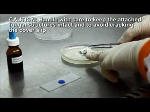
LPCB preparation: Observation of fungal structures from culture.
Serological test:
The monoclonal antibody, EB-A2 used in the commercially available latex agglutination kit to detect galactomannan antigen in sera of patients with penicilliosis. Galactomannan (GM) is a heteropolysaccharide in the cell walls of most Aspergillus and Penicillium species.
Cytological and Histological Examination
The diagnosis of penicilliosis may be suspected or made through examination of cytology or biopsy specimens. Cytology specimens are more readily obtained by less invasive procedures such as FNAC of lymph nodes, sputum cytology, and touch smear of skin. For high-grade fungemia, yeast cells may be seen inside monocytes in peripheral blood smear. The yeast cells may be sparse or abundantly found in histiocytes or extracellularly, and are most readily demonstrated by fungal stains such as periodic acid-Schiff (PAS) and silver methenamine stains. Detection of non-budding yeast cells with characteristic central transverse septum would give a presumptive diagnosis which should be confirmed by microbiological culture.
Molecular test: ITS and/or β-tubulin loci are recommended for the identification of Penicillium species.
Penicillium Growth on SDA,
Morphology of Penicillium,
Macroscopic features-
Microscopic Features-
Pathogenicity
Major diseases are caused by Penicillium species Talaromycosis (formerly Penicilliosis). Penicilliosis is an infection caused by Penicillium marneffei, a dimorphic fungus endemic to Southeast Asia and the southern part of China. It is the 3rd most common opportunistic infection in HIV-positive individuals. Human to human transmission does not occur. Dissemination of infection occurs through the lymphatics or hematogenous. Other than P. marneffei may also cause opportunistic infections leading to mycotic keratitis, otomycosis, and endocarditis.
Laboratory Diagnosis
Specimen: It depends on the nature of the infection site e.g. in the diagnosis of keratitis corneal scrapings (most frequent) or tissue biopsy and skin lesions (either cellulitis or metastatic lesions) while in otitis media ear discharge. Other samples may also bone marrow examination and less reliably from blood cultures be used.
KOH mount: Presence of fungal elements
Fungal culture: To obtain growth of fungi.
LPCB preparation: Observation of fungal structures from culture.
Serological test:
The monoclonal antibody, EB-A2 used in the commercially available latex agglutination kit to detect galactomannan antigen in sera of patients with penicilliosis. Galactomannan (GM) is a heteropolysaccharide in the cell walls of most Aspergillus and Penicillium species.
Cytological and Histological Examination
The diagnosis of penicilliosis may be suspected or made through examination of cytology or biopsy specimens. Cytology specimens are more readily obtained by less invasive procedures such as FNAC of lymph nodes, sputum cytology, and touch smear of skin. For high-grade fungemia, yeast cells may be seen inside monocytes in peripheral blood smear. The yeast cells may be sparse or abundantly found in histiocytes or extracellularly, and are most readily demonstrated by fungal stains such as periodic acid-Schiff (PAS) and silver methenamine stains. Detection of non-budding yeast cells with characteristic central transverse septum would give a presumptive diagnosis which should be confirmed by microbiological culture.
Molecular test: ITS and/or β-tubulin loci are recommended for the identification of Penicillium species.
 0:01:09
 0:00:47
 0:02:31
 0:02:22
 0:01:46
 0:02:12
 0:00:31
 0:02:58
 0:01:16
 0:52:25
 0:00:36
 0:00:07
 0:03:00
 0:01:45
 0:02:37
 0:01:55
 0:02:10
 0:01:10
 0:01:39
 0:03:16
 0:02:01
 0:00:49
 0:05:24
 0:00:11