filmov
tv
Small Bowel Obstruction || Ultrasound || Case 153

Показать описание
Small Bowel Obstruction || Ultrasound || Case 153
Clinical Features:
A middle aged female patient came with
- Abdominal pain
- Abdominal distension
- Nausea & vomiting
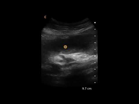
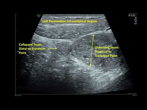
Ultrasound Findings:
- Dilated small intestinal loops with ‘to & fro’ appearance of intraluminal contents.
- Prominence of valvulae conniventes.
- Presence of mural gas.
- Pointy triangular interloop free fluid forming ‘Tanga sign’.
Thank you for watching.
Share with your friends.
Like & Subscribe for more videos.
Don't forget to put your valuable opinions in the Comment box below.
Check the playlist for Congenital Anomaly Lecture videos:
Check the playlist for Imaging Study Lecture videos
Check the playlist for Imaging Study Ultrasound Case videos
Follow us on
#Ultrasound #Radiology #ImagingStudy #Ultrasoundcases #Medical #Imaging #Surgery #Oncology #intestine #emergency #doppler #doctor #isuog #aium #surgeon
Clinical Features:
A middle aged female patient came with
- Abdominal pain
- Abdominal distension
- Nausea & vomiting
Ultrasound Findings:
- Dilated small intestinal loops with ‘to & fro’ appearance of intraluminal contents.
- Prominence of valvulae conniventes.
- Presence of mural gas.
- Pointy triangular interloop free fluid forming ‘Tanga sign’.
Thank you for watching.
Share with your friends.
Like & Subscribe for more videos.
Don't forget to put your valuable opinions in the Comment box below.
Check the playlist for Congenital Anomaly Lecture videos:
Check the playlist for Imaging Study Lecture videos
Check the playlist for Imaging Study Ultrasound Case videos
Follow us on
#Ultrasound #Radiology #ImagingStudy #Ultrasoundcases #Medical #Imaging #Surgery #Oncology #intestine #emergency #doppler #doctor #isuog #aium #surgeon
 0:00:56
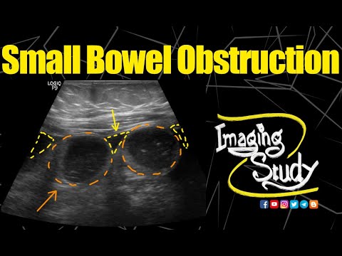
Small Bowel Obstruction - Ultrasound Image Interpretation
 0:08:56
Abdominal Ultrasound Normal Bowel Vs Small Bowel Obstruction Image Appearances | Dilated Bowel USG
 0:01:59
Small Bowel Obstruction || Ultrasound || Case 152
 0:02:35
Small Bowel Obstruction || Ultrasound || Case 153
 0:12:36
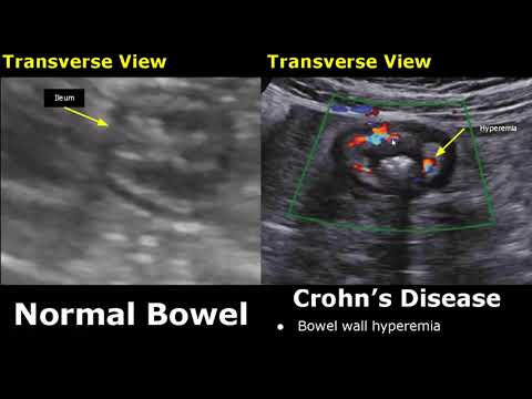
Small Bowel Ultrasound Reporting | How To Write USG Report | Bowel Obstruction, Crohn's Disease
 0:11:38
POCUS in Small Bowel Obstruction
 0:09:54
Small Bowel Obstruction Ultrasound : 10/2019
 0:03:43
Small Bowel Obstruction
 0:02:07
Small bowel obstruction on ultrasound | bowel ultrasound | abdominal ultrasound | acute abdomen
 0:02:19
Small Bowel Obstruction
 0:08:31
Small Bowel Ultrasound Normal Vs Abnormal Image Appearances | Gastrointestinal Tract (GIT) USG
 0:00:38
Small Bowel Obstruction
 0:02:02
Differentiating small bowel from large bowel (specifically ileum vs cecum)
 0:12:06
Understanding Bowel Obstruction
 0:08:33
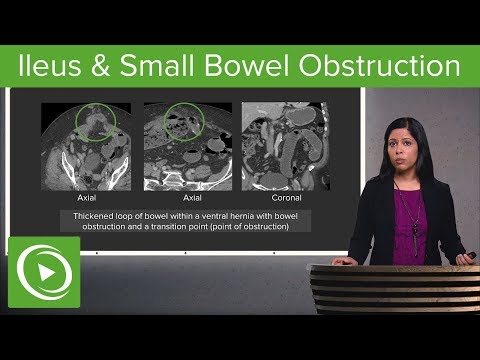
Bowel Obstruction and Ileus: Ileus & Small Bowel Obstruction – Radiology | Lecturio
 0:00:43
🔎 Small vs. Large Bowel: X-Ray Clues! #Radiology #Gastroenterology #USMLE
 0:01:14
Bowel Adhesion Causing Subacute Small Bowel Obstruction-Ultrasound Demonstration of Transition Point
 0:00:45
Small bowel obstruction on abdominal x-ray #radiology #medicalstudent #medicine
 0:00:07
73F,abdominal pain. POCUS: diffuse small bowel dilatation #ApacheUltrasound
 0:01:15
POCUS for Small Bowel Obstruction
 0:05:47
Canine: Intestines
 0:01:20
Large Bowel Obstruction
 0:05:47
Ultrasound Video showing movements of Bowl contents in Intestinal Obstruction.
 0:00:16
Bowel Obstruction - ultrasound
Комментарии