filmov
tv
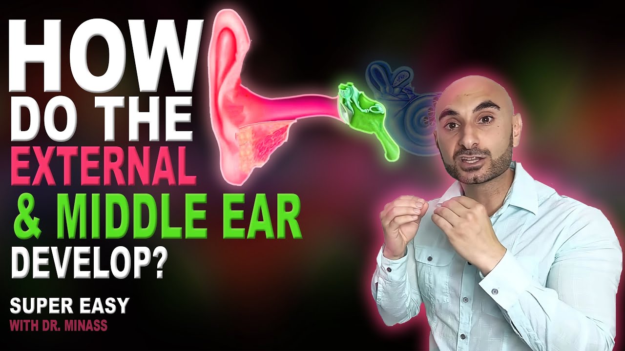
Embryology of the Ear II (Easy to Understand)
Показать описание
The development of the middle and external ear explained in less than 15 minutes.
If you are completely new to embryology and you want to understand it quickly, this should be the first video you watch:
--------------------------------
Recommended Text
--------------------------------
----------------------------------------
Interact With Dr. Minass!
----------------------------------------
Post - Address to:
Minass
Parcel Locker 10106 04448
59 Penshurst Street
Willoughby, NSW
Australia 2068
------------------------------------------------------------------------
SUMMARY OF THE VIDEO FOR YOUR NOTES
------------------------------------------------------------------------
The middle ear
- Tympanic cavity comes from the endoderm, from the first pharyngeal pouch.
- The pouch expands laterally until it touches the first pharyngeal cleft
- The distal part of the pouch is the tubotympanic recess, which widens becoming the primitive tympanic cavity. The proximal part remains narrow and becomes the auditory tube aka eustachian tube
- The malleus and incus come from the first pharyngeal arch
- The stapes comes from the second pharyngeal arch
- Tensor tympani is innervated by the mandibular branch of the trigeminal nerve
- Stapedius is innervated by the facial nerve
The external ear
- External auditory meatus comes from the first pharyngeal cleft
- The solid epithelial meatal plug will dissolve by the seventh month, otherwise the newborn will be deaf
- Th tympanic membrane (eardrum) is made from three layers: ectoderm epithelium, mesoderm middle layer which is connective tissue, and endoderm which is the inner mucous membrane (epithelium).
- The malleus' handle is in contact with the tympanic membrane.
- The auricle develop from 6 swellings called the auricular hillocks.
- Hillocks 1, 2, and 3 come from pharyngeal arch 1
- Hillocks 4, 5, and 6 come from the second arch
- The hillocks will eventually grow and fuse together in a complicated process that I have simplified by just saying "they all fuse together"
- 1 is the tragus, 2 is the helix, 3 is the cymba conca, 4 is the concha, 5 is the anti-helix, and 6 is the anti-tragus - lets keep it at that
 0:14:42
Embryology of the Ear II (Easy to Understand)
 0:05:26
Embryology of ear | development |
 0:16:21
Embryology of the Ear I (Easy to Understand)
 0:19:25
Development of the Ear | Embryology
 0:45:32
3D Embryology of Ear Part 1: Internal Ear - Semicircular Canals - Utricle, Saccule and Cochlea
 0:35:30
3D Middle Ear & External Ear embryology - Impedance matching - Ossicles Embryology
 0:03:31
EMBRYOLOGY OF EAR WITH EASY TRICKS!!
 0:08:05
Development of Ear(Part-2)/Inner Ear/Embryology
 0:24:27
Karl Koehler, PhD, Modeling Inner Ear Development and Disease With Organoids
 0:08:13
Anatomy of the Ear
 0:09:21
Development of Ear (Part-1)/ Embryology
 0:46:48
Special Senses | External & Middle Ear Anatomy
 0:21:19
Special Embryology - Development of the ear
 0:09:20
Embryology : Development of middle ear by Dr.Abhishek kumar
 0:01:00
3D Embryology of Inner Ear in 1 minute #shorts #Ear #Embryology #review #usmle
 0:10:13
Development of the middle ear - The four 'sacci'
 0:08:17
Development of the Face and Palate
 0:05:19
Anatomy - Ear Overview
 0:07:22
Ear Embryo
 0:18:16
3D Embryology of Ear Part 2: Internal Ear Histogenesis of Utricle and Saccule - Perilymphatic Duct
 0:09:35
Embryology: Development of internal ear
 0:36:42
Embryology | Development of Reproductive System
 0:21:50
Special Senses | Inner Ear Anatomy
 0:15:04
Development of ear
Комментарии