filmov
tv
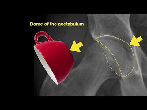
Anatomy of Hip X-rays

Показать описание
___
Lifetime Access to Online Anatomy Course
Foundational Q&A Cards Per Video
Notes and Key Takeaways
Downloadable Documents
Flashcards for Each Course
Weekly Group Tutoring Sessions
Direct Tutoring Sessions
___
Discover A Simplified Approach to Master the Complexity of Anatomy with me, Dr. David Morton ... The Noted Anatomist!
------------------------------------------------------------------
This video tutorial presents the anatomy of hip x-rays:
0:00. Intro to hip x-rays
0:16. Standard hip series for x-rays
0:18. AP view (right hip)
4:19. Lateral view (right hip)
5:34. In-a-Nutshell
5:42. Acknowledgements
 0:06:10
Anatomy of Hip X-rays
 0:11:17
How to read hip x-rays | EASY GUIDE
 0:20:38
Pelvis Anatomy | Radiology anatomy part 1 prep | Pelvic X-ray interpretation
 0:12:02
Pelvis/Hip X-ray interpretation
 0:26:37
Imaging of the Hip: More Radiographic Essentials!
 0:19:51
Hip X-Ray Interpretation: Diagnosing Neck Of Femur Fractures
 0:04:34
Know your hip fractures and the importance of arterial anatomy of the proximal femur
 0:03:06
How To Read An X ray Of Your Hip
 0:00:04
Anatomy of chest Xray #xray #chestxray #motivation #shorts
 0:07:58
Anatomy of Knee X-rays
 0:09:38
Anatomy of Elbow X-rays
 0:12:48
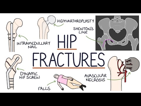
Understanding Hip Fractures and Hip Surgery
 0:07:26
Lower Limb Radiological Anatomy | X-Rays of Hip, Knee Ankle, Foot | For Anatomy Viva
 0:13:33
Pediatric HIP anatomy and most common pathology (radiological anatomy; episode 14)
 0:04:18
X-ray of Hip Joint | Pelvis | Clinical Anatomy | By Dr.(Prof) Sibani Mazumdar
 0:18:29
Hip X-Rays in ED
 0:04:38
anatomy of hip x-rays
 0:57:30
SSR Resident Education Club - Approach to Hip MRI
 0:09:27
Anatomy of Hand X-rays_Revised
 0:17:18
Radiological Anatomy of Lower Limb
 0:04:51
Hip Joint - Part 4 - Normal Anatomy - Anterior and Posterior Columns of the Acetabulum
 0:01:12
Where is the Sacroiliac Joint? Anatomy of the Sacroiliac Joint
 0:05:23
Hip with Pelvis X-ray Positioning video
 0:17:50
Wrist bones and alignment | Radiology anatomy part 1 prep | Wrist X-ray interpretation
Комментарии