filmov
tv
Brain MRI - Seizure search pattern

Показать описание
Many times when patients have a history of seizures, they undergo a workup including a physical exam, detailed EEG analysis, and finally brain MRI to try to identify any potential structural causes of seizures. In this video, Dr. Michael Hoch walks us through his approach to a brain MRI to maximize your sensitivity for finding abnormalities.
The lecture is divided as follows:
00:00 - Introduction
01:53 - T1-MPRAGE Sagittal
03:43 - T1-MPRAGE Coronal
05:26 - T1-MPRAGE Axial
06:04 - FLAIR
07:39 - GRE
07:54 - Hippocampus
11:11 - Summary
Dr. Hoch suggests a 4-step approach using the mnemonic “3-2-1 go to the hippocampus”. In this way, he divides his search into more digestible parts.
“3” indicates the 3 planes that you have in a non-contrast T1 weighted MP-RAGE MRI. On this you should focus on the cortex, particularly at the 3 poles, the frontal, temporal, and occipital poles.
“2” indicates the 2 planes of FLAIR and 2 window settings you should use. You should review FLAIR images in both the coronal and axial planes. You should also use a window that is normal and a window that is narrow, or aggressive, to highlight lesions, particularly in the cortex, which are hard to see.
“1” indicates the single plane of blood sensitive imaging, either GRE or SWI, which can often see areas of prior hemorrhage or cavernous malformations.
“Go” to the hippocampus last to look for signs of mesial temporal sclerosis, which is manifested as a small hippocampus with loss of internal architecture and abnormal T2/FLAIR hyperintensity. This can be either from primary epilepsy or secondary to another lesion.
 0:11:51
Brain MRI - Seizure search pattern
 0:03:48
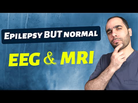
Epilepsy With Normal EEG and MRI Brain?
 0:01:53
The Difference between Seizures and Epilepsy
 0:01:07
What Happens in Your Brain During a Seizure | WebMD
 0:04:35
Will brain damage show on MRI?
 0:01:32
What to expect during your child's MRI?
 0:00:13
Hippocampal sulcal remnant cyst#Epilepsy imaging #MRI epilepsy#Seizures
 0:54:29
The Seizing Brain (part 2) - Seizure-induced MRI abnormalities
 1:25:20
The Seizing Brain - Seizure-induced imaging abnormalities on perfusion-CT and MRI.
 0:01:23
Absence Seizures: EEG Findings
 0:02:06
High-tech imaging allowed doctors to find area causing seizures
 0:04:37
What happens during an MRI examination?
 0:01:58
Seizures: What Causes Seizures? Symptoms and when you need to see a Doctor.
 0:23:52
Seizure Imaging Part 1 | Health4TheWorld Academy
 0:14:22
Seizure Imaging Part 3 | Health4TheWorld Academy
 0:04:09
Mri Brain Epilepsy protocol sequences @ Seizure protocol Identification
 0:00:24
IR Sequence, Seizure protocol #viral #mri #youtubeshorts
 0:11:10
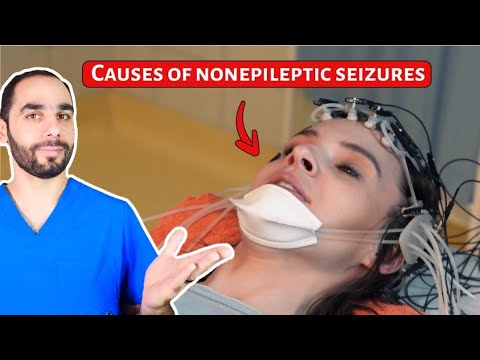
Causes of Nonepileptic Seizures (PNES). Hint, NOT Stress!
 0:01:59
High Powered Imaging Solves Teen Seizure Mystery PKG (HD)
 0:19:08
Beyond Search & Seizure | Jeffrey Rosen | TEDxPhiladelphia
 0:02:21
Colorado Mother Prepares For Brain Surgery To Find Source Of Epileptic Seizures
 0:03:42
Neuroscience: Epilepsy: Preventing Seizures with Brain Stimulations
 0:01:04
MRI Brain seizure Protocols/ Epilepsy Protocol #mri #ctscan #radiology
 0:02:26
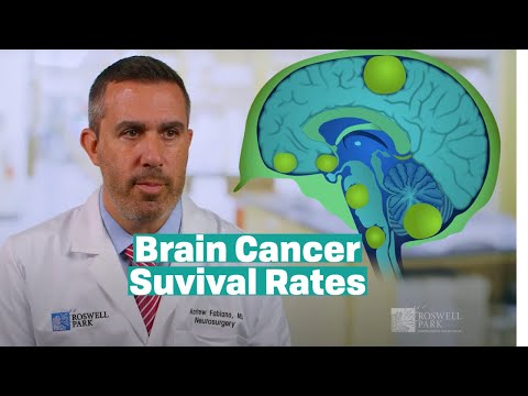
Understanding Brain Tumor Survival Rates
Комментарии