filmov
tv
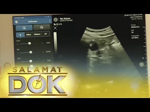
Which is better: abdominal ultrasound or TVS? #ultrasound #baby #pregnant #pregnancyjourney #momtobe

Показать описание
What is ultrasound TVS scan?
Transvaginal sonography also known as TVS scan or USG TVS or Endovaginal ultrasound is a type of ultrasound imaging that is used for examination of female pelvic organs or reproductive system including uterus, cervix, ovary and fallopian tubes.
It is a rapid, non-invasive and usually painless technique that enable more detailed visualization of pelvic organs as compared to transabdominal ultrasonography. The main difference between Transabdominal ultrasound and a TVS scan is the positioning of probe/transducer. In transabdominal ultrasonography the probe/transducer is kept on the abdominal surface where as in TVS scan the transducer is kept inside your vagina.
Chronic pelvic pain
Pelvic infections
Pelvic inflammatory disease (PID)
Abnormal or excessive vaginal bleeding
Post-menopausal bleeding
Ovarian cysts
Uterine Polyp
To identify any abnormality in the shape, size, location and blood supply of uterus including endometrium, fallopian tubes and ovaries.
To diagnose Ectopic pregnancy
Pelvic masses or cysts
Any abnormal growth such as cancers of uterus, cervix or ovaries
Uterine Fibroids
Infertility
Abortions or miscarriage
To confirm pregnancy in initial stages and identify pregnancy associated conditions.
Pre-operative evaluation before pelvic surgery.
Follow up or monitoring of patients post-treatment.
How do I prepare for USG TVS test?
Follow these simple steps if you are going for transvaginal Ultrasonography scan-
Before the scan begins you need to remove Tampon, ornaments and clothes.
You will be then asked to lie on the examination table.
A technician may measure blood pressure if TVS color doppler is scheduled.
Your Sonographer/Obstetrician will then apply gel/lubricant over a condom which is placed over the transducer for the smooth insertion of probe into your vagina.
Then a transducer/probe will be inserted inside your vagina which sends high frequency sound waves to the pelvic organs. These sound waves are reflected back and are recorded by the ultrasound machine and converted into images. These images will be interpretated to evaluate your condition.
At the end of procedure, tissue paper will be provided to clean the gel/lubricant .
You will be allowed to leave once the imaging is over.
What happens after Transvaginal ultrasound?
TVS Ultrasound/USG test usually takes about 15-30 minutes. However, it may lasts longer depending the severity of condition and the associated co-morbid conditions.
You will be allowed to leave immediately after the scan.
After the procedure got over, your Sonographer will analyze the result and make a diagnosis of your condition.
You will get the reports on the same day.
These reports will be evaluated by your Obstetrician to make a proper management plan for your condition.
TVS scan is usually a safe procedure with no side effects. However, some women may feel a bit discomfort during insertion of transducer into vagina.
A prenatal or pregnancy ultrasound uses sound waves to create a picture of your baby on a screen. Pregnancy care providers use it to check on the health of your baby and detect certain pregnancy complications. Most people have two ultrasounds during pregnancy, but you may have more if your provider feels it’s medically necessary.
What is an ultrasound in pregnancy?
A prenatal ultrasound (or sonogram) is a test during pregnancy that checks on the health and development of your baby. An obstetrician, nurse midwife or ultrasound technician (sonographer) performs ultrasounds during pregnancy for many reasons. Sometimes ultrasounds occur to check on your baby and make sure they’re growing properly. Other times your pregnancy care provider orders an ultrasound after they detect a problem.
During an ultrasound, sound waves are sent through your abdomen or vagina by a device called a transducer. The sound waves bounce off structures inside your body, including your baby and your reproductive organs. Then, the sound waves transform into images that your provider can see on a screen. It doesn’t use radiation, like X-rays, to see your baby.
#pregnancy #pregnant #baby #newborn #motherhood #maternity #love #babygirl #momtobe #babyboy #momlife #family #babyshower #birth #babybump #mom #weekspregnant #babies #maternityphotography #postpartum #photography #pregnantbelly #mumtobe #breastfeeding #maternityshoot #parenting #pregnancyannouncement #mama #infertility #bebe #fertility #health #doula #ivf #kids #babylove #newbornphotography #pregnantlife #newmom #schwanger #photooftheday #preggo #embarazo #womenshealth #grossesse #mother #mommytobe #pregnancyphotoshoot #pregnancyjourney #instagood #pregnancylife #mumlife #babyontheway #childbirth #weeks #parenthood #happy #expecting #maternityfashion #cute #tvsultrasound #ultrasound #sonography #ultrasoundscans #newlyweds #afterbirth #pregnancyjourney
 0:03:27
What Is An Abdominal Ultrasound?
 0:04:30
What to Expect During an Abdominal Ultrasound
 0:03:03
Abdominal Ultrasound - Basics of Evaluating the Liver
 0:12:02
US Abdomen Complete Protocol
 0:00:41
Which is better: abdominal ultrasound or TVS? #ultrasound #baby #pregnant #pregnancyjourney #momtobe
 0:04:40
10 things to know before you go for a Ultrasound Scan
 0:02:59
How to perform an ultrasound exam of the pancreas
 0:02:19
What to Expect From a Female Pelvic Ultrasound Exam
 1:04:33
AUDIOBOOK / Abdominal Migraine: Recognizing Symptoms, Diagnosis, and Effective Treatments
 1:27:14
Abdominal Ultrasound Normal Vs Abnormal Images | Liver, Gallbladder, Pancreas, Kidney, Hernia USG
 0:00:54
How can I prepare for my Abdominal or Pelvic Ultrasound?
 0:13:15
Abdominal Ultrasound: Guidelines for best reporting
 0:01:04
What are the differences between a transvaginal ultrasound and an abdominal ultrasound?
 0:08:11
Liver Ultrasound Probe Positioning | Transducer Placement For Liver Scanning | Abdominal USG
 0:04:36
Salamat Dok: Common diseases found using an ultrasound of the whole abdomen for men
 0:17:01
Abdominal Ultrasound BachelorClass - Your introduction to abdominal ultrasound
 0:13:22
Introduction to the interpretation of Abdominal Ultrasound
 0:05:36
How to ultrasound the liver
 0:00:26
Standardized Ultrasound Abdomen Protocol at IDC
 0:19:57
The evaluation of diagnostic usefulness of abdominal ultrasound in dogs with signs of pancreatitis
 0:00:43
How different abdominal organs look on ultrasound imaging
 0:02:26
Simple, Effective Abdominal Ultrasound at Northwestern Medicine
![[US] Abdominal Ultrasound](https://i.ytimg.com/vi/tUA3mwzSmdk/hqdefault.jpg) 0:11:31
0:11:31
[US] Abdominal Ultrasound | Search Pattern
 0:04:09
Point of Care Ultrasound of the Abdominal Aorta - AMBOSS Video
Комментарии