filmov
tv
Development Of Maxilla - Embryology

Показать описание
#Development of the maxilla
Maxilla is an irregularly shaped paired bone and is formed by the union of two maxillary bones at the intermaxillary suture. It forms the dominant portion of the face. The two main parts maxilla has is the body and the processes. The body occupies the maxillary sinus viewed here from an internal view of a single maxillary bone. The maxillary sinus is pea-sized at birth and gradually increase in size with increasing age. The processes are the frontal, zygomatic, alveolar and palatine process. The frontal process articulates with the frontal bone, the zygomatic process unites with the zygomatic bone, the alveolar process, includes tooth sockets of upper teeth and the palatine process joins with the palatine bone and forms the anterior two/thirds of the palate.
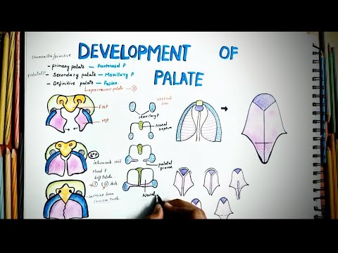
The development of the maxilla arises from the first pharyngeal arch called the mandibular arch. The mandibular arch is one of the five pairs of pharyngeal arches present at the neck region of the embryo. Out of these five arches the first arch named the mandibular arch soon gives off a division by the end of week 4. This very first division of the mandibular arch is called the maxillary prominence. The maxillary prominence will eventually give rise to the maxillary bone through the process of intramembranous bone formation.
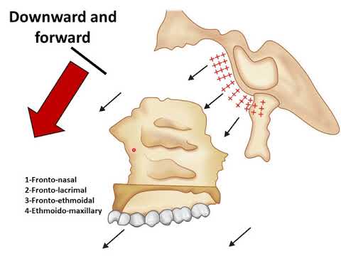
No arch cartilage is associated with the formation of the maxillary bone. The center of ossification from which the bone formation begins actually has a close association with the cartilage of the nasal capsule. The center of ossification appears in the angle between the infraorbital nerve and the anterosuperior alveolar nerve. Both of these nerves are divisions of maxillary branch of fifth cranial nerve or trigeminal nerve. The maxillary teeth are innervated by three superior alveolar nerve branches. That is anterior superior, middle superior and posterior superior alveolar nerves. From the center of ossification the bone formation spreads in so many directions to form the complex bone of maxilla. Ossification spreads posteriorly below the orbit and towards the developing zygoma. Anteriorly, it spreads towards the future incisor teeth. Superiorly to form the frontal process and then it also spreads towards the palatine process to form the hard palate and towards the main body of the maxilla. Later on a secondary cartilage, contributes to the development of the maxilla. This secondary cartilage is present at the zygomatic process of the maxilla which persists here for a short time and it adds considerably to the development of the maxilla.
Video (diagrams+editing) & Voiceover:
Dr.Maryam Kazman
Become a Patreon and Get Exclusive Rewards:
Join me on facebook:
Maxilla is an irregularly shaped paired bone and is formed by the union of two maxillary bones at the intermaxillary suture. It forms the dominant portion of the face. The two main parts maxilla has is the body and the processes. The body occupies the maxillary sinus viewed here from an internal view of a single maxillary bone. The maxillary sinus is pea-sized at birth and gradually increase in size with increasing age. The processes are the frontal, zygomatic, alveolar and palatine process. The frontal process articulates with the frontal bone, the zygomatic process unites with the zygomatic bone, the alveolar process, includes tooth sockets of upper teeth and the palatine process joins with the palatine bone and forms the anterior two/thirds of the palate.
The development of the maxilla arises from the first pharyngeal arch called the mandibular arch. The mandibular arch is one of the five pairs of pharyngeal arches present at the neck region of the embryo. Out of these five arches the first arch named the mandibular arch soon gives off a division by the end of week 4. This very first division of the mandibular arch is called the maxillary prominence. The maxillary prominence will eventually give rise to the maxillary bone through the process of intramembranous bone formation.
No arch cartilage is associated with the formation of the maxillary bone. The center of ossification from which the bone formation begins actually has a close association with the cartilage of the nasal capsule. The center of ossification appears in the angle between the infraorbital nerve and the anterosuperior alveolar nerve. Both of these nerves are divisions of maxillary branch of fifth cranial nerve or trigeminal nerve. The maxillary teeth are innervated by three superior alveolar nerve branches. That is anterior superior, middle superior and posterior superior alveolar nerves. From the center of ossification the bone formation spreads in so many directions to form the complex bone of maxilla. Ossification spreads posteriorly below the orbit and towards the developing zygoma. Anteriorly, it spreads towards the future incisor teeth. Superiorly to form the frontal process and then it also spreads towards the palatine process to form the hard palate and towards the main body of the maxilla. Later on a secondary cartilage, contributes to the development of the maxilla. This secondary cartilage is present at the zygomatic process of the maxilla which persists here for a short time and it adds considerably to the development of the maxilla.
Video (diagrams+editing) & Voiceover:
Dr.Maryam Kazman
Become a Patreon and Get Exclusive Rewards:
Join me on facebook:
 0:03:48
Development Of Maxilla - Embryology
 0:22:40
PRENATAL DEVELOPMENT OF MAXILLA II GROWTH OF MAXILLA II EMBRYOLOGY II ORTHODONTICS
 0:08:17
Development of the Face and Palate
 0:10:21
MAXILLA (Growth and Development) - Orthodontics & Dentofacial Orthopaedics
 0:08:45
Prenatal growth : maxilla & mandible | development of palate
 0:04:34
DPES EarlyEmbryonicFacialDevelopment
 0:12:57
Development of Mandible | Anatomy and Embryology for Medical Students
 0:22:27
POST-NATAL GROWTH OF MAXILLA II MECHANISMS AND CLINICAL SIGNIFICANCE II EMBRYOLOGY II ORTHODONTICS
 0:29:49
Growth of maxilla
 0:14:08
DEVELOPMENT OF MANDIBLE II PRENATAL GROWTH OF MANDIBLE II EMBRYOLOGY II ORTHODONTICS
 0:31:51
Orthodontics | Growth & Development | INBDE, ADAT
 0:04:01
Development of the Mandible - Embryology [ Learn it in the most SIMPLE way]
 0:02:33
Prenatal Growth of Maxilla | Concised | Orthodontics
 0:01:28
How does the mandible grow?
 0:06:53
Prenatal Development of maxilla and palate | Embryology | Anatomy
 0:14:15
growth of maxilla
 1:22:25
Growth of Maxilla and mandible
 0:05:42
Prenatal and Postnatal growth of Maxilla | Growth and Development Part 3 | Pediatric Dentistry #mds
 0:29:30
Growth of Maxilla
 0:07:18
#maxilla #maxillabone #developmentofmaxilla maxilla bone||maxilla in 3D||development of maxilla
 0:00:25
Animation - Maxilla 3D Model v3
 0:43:00
Development of Maxilla & Mandible | Orthodontics | Yenepoya Dental College
 0:09:42
Development Of Palate || Embryology || Easy Explaination || Cleft Palate || Defective Development
 0:10:11
Development of Mandible and Maxilla
Комментарии