filmov
tv
Internal jugular veins anatomy|jugular veins anatomy #madicalhands #cvp #central line #video #viral

Показать описание
hi friends please like 👍 & subscribe ☺️👆my channel ,❤️🥰🎸🙏
#viral #shorts ,#short ,#shortvideo ,#subscribe
#video
#viralvideo
#viralshorts
#views
#video
#shortvideo #viral #viralvideo #viralshort #shorts #Thalapathy #Vijay #trending #tiktok #trendingshorts #bollywood #trendingvideo
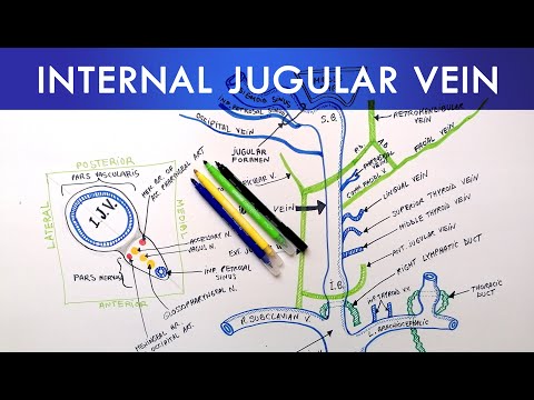
The internal jugular veins are major blood vessels that play a crucial role in draining blood from the head, neck, and brain. Here's an overview of their anatomy:
Location: The internal jugular veins are located on either side of the neck, running parallel to the carotid arteries. They are deep to the sternocleidomastoid muscle, which is a large muscle on the side of the neck.
Course: Each internal jugular vein begins at the jugular foramen, which is an opening at the base of the skull. From there, it descends down the neck, passing through the carotid sheath, which is a fibrous connective tissue structure containing the common carotid artery, the internal carotid artery, and the vagus nerve.
Tributaries: Along its course, the internal jugular vein receives blood from various tributaries. These include the facial vein, lingual vein, pharyngeal veins, superior and middle thyroid veins, occipital vein, and the posterior auricular vein. The internal jugular vein also receives blood from the brain through the dural venous sinuses, which drain into it at the base of the skull.
Valves: The internal jugular veins are equipped with one-way valves that help maintain the unidirectional flow of blood. These valves prevent backflow and ensure efficient drainage of blood toward the heart.
Termination: Near the root of the neck, the internal jugular vein merges with the subclavian vein to form the brachiocephalic vein. The brachiocephalic vein then joins the other side's brachiocephalic vein to form the superior vena cava, which is the large vein that returns blood from the upper body to the heart.
Function: The internal jugular veins are responsible for draining deoxygenated blood from the brain, face, neck, and many structures within the head. They play a crucial role in returning this blood to the heart for oxygenation and systemic circulation.
Clinical Significance: The internal jugular veins are commonly used for central venous access during medical procedures, such as the placement of central venous catheters or for obtaining blood samples. They can also be used for monitoring central venous pressure, which provides information about the heart's function and fluid status. Additionally, abnormalities in the internal jugular veins can be associated with certain medical conditions, such as thrombosis (clot formation) or stenosis (narrowing).
For internal jugular vein CVL (central approach), the needle should be inserted at the triangle's apex formed by the sternocleidomastoid muscle's two heads above the medial clavicle and is usually 5 cm superior to the clavicle. The needle should be inserted at 30 to 45 degrees into the skin.
#viral #shorts ,#short ,#shortvideo ,#subscribe
#video
#viralvideo
#viralshorts
#views
#video
#shortvideo #viral #viralvideo #viralshort #shorts #Thalapathy #Vijay #trending #tiktok #trendingshorts #bollywood #trendingvideo
The internal jugular veins are major blood vessels that play a crucial role in draining blood from the head, neck, and brain. Here's an overview of their anatomy:
Location: The internal jugular veins are located on either side of the neck, running parallel to the carotid arteries. They are deep to the sternocleidomastoid muscle, which is a large muscle on the side of the neck.
Course: Each internal jugular vein begins at the jugular foramen, which is an opening at the base of the skull. From there, it descends down the neck, passing through the carotid sheath, which is a fibrous connective tissue structure containing the common carotid artery, the internal carotid artery, and the vagus nerve.
Tributaries: Along its course, the internal jugular vein receives blood from various tributaries. These include the facial vein, lingual vein, pharyngeal veins, superior and middle thyroid veins, occipital vein, and the posterior auricular vein. The internal jugular vein also receives blood from the brain through the dural venous sinuses, which drain into it at the base of the skull.
Valves: The internal jugular veins are equipped with one-way valves that help maintain the unidirectional flow of blood. These valves prevent backflow and ensure efficient drainage of blood toward the heart.
Termination: Near the root of the neck, the internal jugular vein merges with the subclavian vein to form the brachiocephalic vein. The brachiocephalic vein then joins the other side's brachiocephalic vein to form the superior vena cava, which is the large vein that returns blood from the upper body to the heart.
Function: The internal jugular veins are responsible for draining deoxygenated blood from the brain, face, neck, and many structures within the head. They play a crucial role in returning this blood to the heart for oxygenation and systemic circulation.
Clinical Significance: The internal jugular veins are commonly used for central venous access during medical procedures, such as the placement of central venous catheters or for obtaining blood samples. They can also be used for monitoring central venous pressure, which provides information about the heart's function and fluid status. Additionally, abnormalities in the internal jugular veins can be associated with certain medical conditions, such as thrombosis (clot formation) or stenosis (narrowing).
For internal jugular vein CVL (central approach), the needle should be inserted at the triangle's apex formed by the sternocleidomastoid muscle's two heads above the medial clavicle and is usually 5 cm superior to the clavicle. The needle should be inserted at 30 to 45 degrees into the skin.
 0:01:18
Internal jugular veins anatomy|jugular veins anatomy #madicalhands #cvp #central line #video #viral
 0:04:12
Internal Jugular Vein | Anatomy Tutorial
 0:14:55
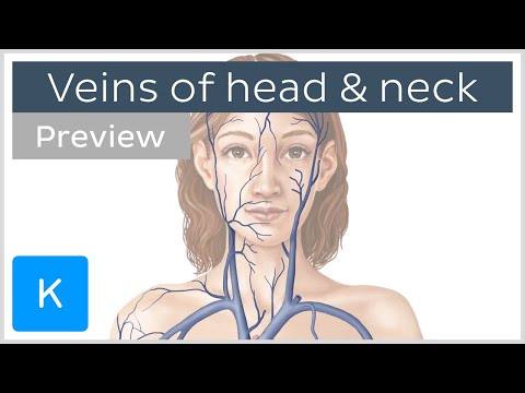
FULL VIDEO: Main veins of the head and neck - Human Anatomy | Kenhub
 0:02:06
Internal Jugular Vein | Course | Tributaries
 0:01:02
internal jugular vein anatomy model lab
 0:05:48
IJV - What's the magic in measuring the Internal Jugular Vein?
 0:10:29
Veins of the Head and Neck (EASY Scheme) + Mnemonics & Quiz
 0:09:05
Internal Jugular Vein - Commencement | Termination | Relations | Tributaries | Applied Anatomy
 0:11:25
Understanding Jugular Venous Pressure (JVP)
 0:08:23
Blind new technique for IJV cannulation
 0:08:23
Veins of the Neck | Subclavian | Jugular veins | Functions | Clinical significance | Anatomy
 0:08:52
Internal jugular vein catheterization |Sanjay Das ( MT Dialysis )|R.G Kar medical college
 0:00:45
Regular Carotid Pulsation and Discernible Internal Jugular Venous Pulse
 0:01:31
EJV (External Jugular vein ) Cannulation 💉
 0:01:50
Cardiology - Assessing the Jugular Venous Pulse
 0:00:12
Blood vessels of the head and neck
 0:05:08
Anatomy Tutorial - Veins of the Head and Neck
 0:07:27
Anterior Neck - Veins
 0:15:05
Circulatory System | Veins of the Head & Neck | Flow Chart
 0:03:39
Veins of the head and neck (preview) - Human Anatomy | Kenhub
 0:02:15
Examination of JVP in Internal Jugular Vein, Dept. Of Medicine, JNMC , Wardha
 0:00:33
internal jugular vein surface marking|| surface anatomy of internal jugular vein
 0:17:47
INTERNAL JUGULAR VEIN - ANATOMY SERIES
 0:01:31
POCUS for Internal Jugular Vein
Комментарии