filmov
tv
What do bacteria look like under a light vs electron microscope?

Показать описание
Check out how our scientists prepare bacterial samples to look at them under light and electron microscopes. The reagents used and the protocols followed are different - check out what these are and what purpose do they serve. And finally see how bacteria looks like under a microscope.
Typo at 0.25: Take 0.01 ml (not 0.1ml) of overnight culture and add it to 1ml of fresh medium to dilute it 100 times.
Typo at 0.25: Take 0.01 ml (not 0.1ml) of overnight culture and add it to 1ml of fresh medium to dilute it 100 times.
 0:00:43
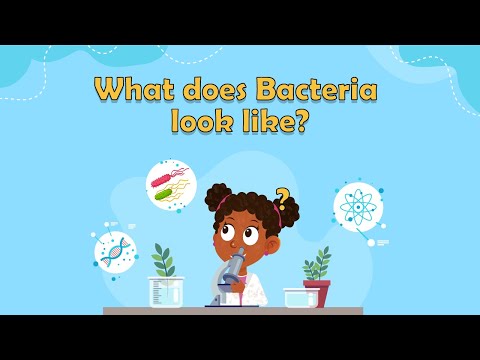
What does Bacteria look like? | What is bacteria for kids | Bacteria facts for kids | Bacteria Cell
 0:09:40
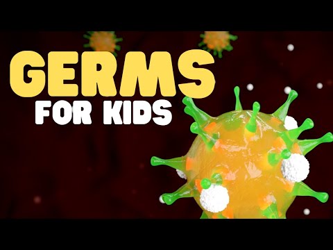
Germs for Kids | Learn all about bacteria, viruses, fungi, and protozoa
 0:00:38
Vinegar VS Bacteria under the microscope!
 0:00:32
WHAT THINGS ACTUALLY LOOK LIKE UNDER A MICROSCOPE
 0:00:35
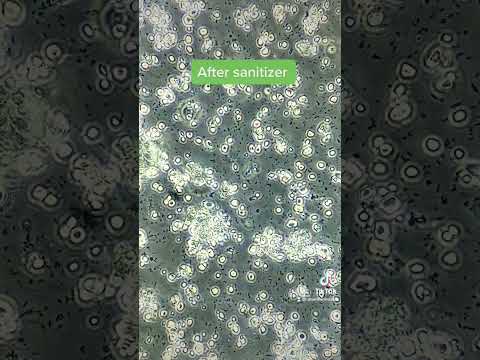
Bacteria VS hand sanitizer under the microscope!
 0:00:59
White Blood Cell Fights GIANT GERM!
 0:00:39
Tap water under the microscope! (You will be surprised!)
 0:00:13
Virus and bacteria under electron microscope
 1:47:37
What Do Aliens Look Like? Exploring the Evolutionary Scenarios of Extraterrestrial Life
 0:02:27
This is what the bacteria on your hands looks like under a microscope
 0:03:56
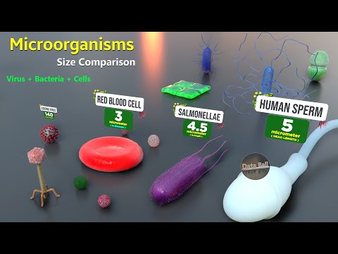
How Microorganisms looks under the microscope | Virus Size | Bacteria size | Antibodies size
 0:00:41
Bacteria 3D Animation
 0:00:41
Bacteria VS Tequila alcohol under the microscope!
 0:00:56
Genetically Modifying Bacteria Speed Run
 0:00:13
Hand Under Microscope | TULO - Microscope #shorts #microscope #hand #underthemicroscope #bacteria
 0:00:38
Bacteria filmed 'hiding' from antibiotic
 0:04:20
What do bacteria look like under a light vs electron microscope?
 0:00:58
Something weird happens when you add ANTIBIOTICS to BACTERIA under the microscope!
 0:00:16
1 billion bacteria in your tooth plaque | TULO - Microscope #shorts #toothpaste #bacteria
 0:00:32
Banana Under Microscope 🍌#microscopeview #experiment #banana #bacteria #microscope #science #shorts...
 0:00:25
Microscopic Highway of Bacteria Under Microscope #science #microbiology #microscope
 0:00:12
Nose rust under the microscope | TULO - Microscope #shorts #microscope #bacteria #nose
 0:00:42
Mouthwash VS Bacteria under the microscope!
 0:00:21
Bacteriophage 3D Animation|| Structure of Bacteriophage|| How Bacteriophage infect Bacteria?
Комментарии