filmov
tv
Flow Cytometry Introduction - Malte Paulsen (EMBL)

Показать описание
This video provides an excellent introduction to flow cytometry. Dr. Malte Paulsen covers the basic principles of the technique including fluidics, optics and data display.
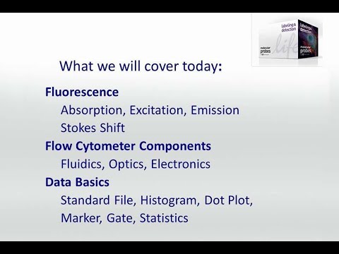
Talk Overview:
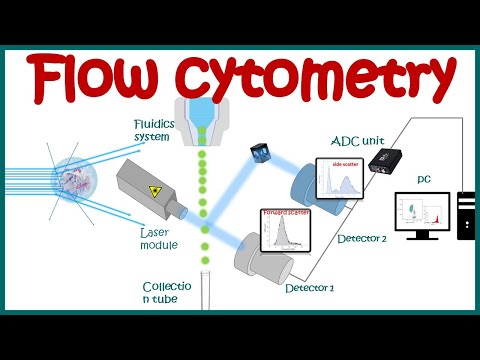
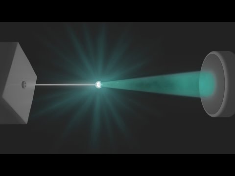
Dr. Malte Paulsen gives an introduction to flow cytometry with an excellent explanation of the basic principles governing the technique. He explains how fluid flow is used to focus a sample in a laser beam. Light from the laser is scattered by cells in the sample and the degree of scatter provides information about the cell’s optical density and other characteristics. In conventional flow cytometry, lasers are used primarily to excite fluorescent antibodies bound to specific cell types. A detector with different filters allows specific wavelengths to be dissected from the overall fluorescence. This signal can then be displayed in ways that provide the most information about the cell type of interest.
Speaker Biography:
Dr. Malte Paulsen has been Head of the Flow Cytometry Core Facility at EMBL since 2015. Prior to joining EMBL, Paulsen was Head of the flow cytometry facilities first at Institute for Molecular Biology (IMB) in Mainz, and later at the National Heart and Lung Institute, Imperial College, London. Paulsen received his PhD from the German Cancer Research Center and the University of Heidelberg in 2011.
 0:33:55
Flow Cytometry Introduction - Malte Paulsen (EMBL)
 0:08:50
Flow cytometry : basic principles | What the use of flow cytometry ? | Cell sorting by FACS
 0:06:18
Invitrogen Attune NxT Flow Cytometer Demo Video
 0:04:35
Flow Cytometry Animation
 0:13:24
Flow Cytometry Analysis
 0:04:42
Flow Cytometry
 0:12:05
Molecular Probes Tutorial Series—Introduction to Flow Cytometry
 0:06:35
Flow Cytometry – Liliana Carvalho
 0:05:13
Intro to Flow Cytometry
 0:10:57
Flow cytometry introduction and troubleshooting
 0:17:41
Molecular Probes Tutorial Series—Analyzing Flow Cytometry Data
 0:01:12
How to Choose Flow Cytometry Antibodies
 0:06:02
What is FLOW CYTOMETRY (Intro to Flow Cytometry - Episode 1)
 0:14:47
Flow cytometry introduction and sample preparation
 0:26:09
Flow Cytometry Introduction
 0:53:47
Webinar: Making polychromatic flow cytometry easy
 0:38:50
Mass Cytometry Introduction - Susanne Heck (NIHR BRC)
 0:56:07
Basics of flow cytometry, Part I: Gating and data analysis
 0:00:14
What is Flow Cytometry? #pathagonia #flowcytometry #hemepath #heme #lymphoma #hematology #lymphomas
 0:02:06
Compensation in Clinical Flow Cytometry
 0:07:41
Robotic Automation for Flow Cytometry
 0:02:19
MACS Facts: Autolabeling with the MACSQuant Flow Cytometer
 0:01:10
Attune NxT: The Band of Acoustic Cytometry
 0:42:37
Flow cytometry basics: Overcome technical challenges [WEBINAR]
Комментарии