filmov
tv
Webinar 1: Breast Cancer Ultrasound Imaging

Показать описание
Webinar 1: 31 May 2021
Featured Topic: Breast Cancer Ultrasound Imaging
Current ultrasound imaging techniques use only part of the information enclosed in the recorded high-frequency sound waves limiting the quality of the information present in the reconstructed image. Advanced ultrasound imaging methods, known as full waveform inversion, use all available information enclosed in the recorded field – including multiple scattering, dispersion, and diffraction effects - to improve the image quality and give accurate quantitative information about the tissue parameters. Full-wave inversion (FWI) may be implemented in either the time or the frequency domain but determining which method should be used is not simple. In this webinar, we will discuss two different non-linear FWI imaging methods: time-domain inversion (TDI) and frequency-domain contrast source inversion (CSI). The methods were tested on the reconstruction of the same synthetic data; a 2-D scan from a circular transducer array enclosing a cancerous breast model. Both methods were evaluated in noise-free and noisy scenarios. Also, the reciprocity of sources and receivers was evaluated as well as the computational complexity of the methods.
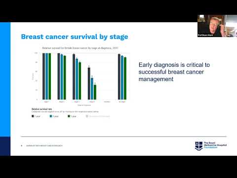
Lymphedema in breast cancer survivors is a chronic condition that involves the upper limb homolateral to therapeutic procedures related to breast cancer treatment. Currently, lymphedema is typically diagnosed clinically based on a patient’s history and characteristic physical findings. Recent advancements provide opportunities for new imaging techniques to assist in the diagnosis of lymphedema based on anatomical changes and guide therapeutic intervention. Diagnostic ultrasound is a potential tool to see, evaluate and quantify the composition of upper limb tissue and also blood circulation in lymphedema.
Featured Topic: Breast Cancer Ultrasound Imaging
Current ultrasound imaging techniques use only part of the information enclosed in the recorded high-frequency sound waves limiting the quality of the information present in the reconstructed image. Advanced ultrasound imaging methods, known as full waveform inversion, use all available information enclosed in the recorded field – including multiple scattering, dispersion, and diffraction effects - to improve the image quality and give accurate quantitative information about the tissue parameters. Full-wave inversion (FWI) may be implemented in either the time or the frequency domain but determining which method should be used is not simple. In this webinar, we will discuss two different non-linear FWI imaging methods: time-domain inversion (TDI) and frequency-domain contrast source inversion (CSI). The methods were tested on the reconstruction of the same synthetic data; a 2-D scan from a circular transducer array enclosing a cancerous breast model. Both methods were evaluated in noise-free and noisy scenarios. Also, the reciprocity of sources and receivers was evaluated as well as the computational complexity of the methods.
Lymphedema in breast cancer survivors is a chronic condition that involves the upper limb homolateral to therapeutic procedures related to breast cancer treatment. Currently, lymphedema is typically diagnosed clinically based on a patient’s history and characteristic physical findings. Recent advancements provide opportunities for new imaging techniques to assist in the diagnosis of lymphedema based on anatomical changes and guide therapeutic intervention. Diagnostic ultrasound is a potential tool to see, evaluate and quantify the composition of upper limb tissue and also blood circulation in lymphedema.
 1:04:37
 0:45:57
 0:59:46
 1:00:46
 1:11:15
 0:56:18
 0:29:41
 1:12:39
 0:49:08
 0:18:52
 0:56:50
 1:04:03
 1:06:20
 0:59:53
 1:06:01
 0:36:30
 1:07:40
 0:54:02
 0:54:30
 1:14:23
 0:58:50
 1:00:31
 1:27:29
 0:38:06