filmov
tv
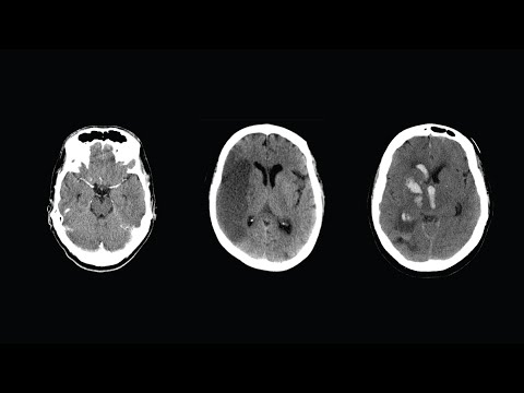
CT Scan Brain Normal Vs Ischemic Stroke Images | Non-Contrast Hyperacute/Acute/Chronic Infarction

Показать описание
CT Scan Brain Normal Vs Ischemic Stroke Images | Non-Contrast Hyperacute/Acute/Chronic Infarction
*Cases:
Intro - 0:00
Ischemic Stroke- Immediate (Hyperdense MCA Sign) - 0:12
Hyperacute - 4:29
Acute - 7:48
Subacute - 9:35
Chronic - 10:56
Hyperacute:
Loss of Gray-white matter differentiation
Hypodense cortex
Gyral effacement - Flattening of gyri
Disappearance of sulci
Acute:
Hypodense areas
Cytotoxic cerebral edema (Swelling)
Mass effect
Midline Shift
Sulcal Disappearance
Subacute:
Reduction of swelling
CT Fogging Effect- Hypodense areas become isodense
Normal Appearance
Cortical petechial hyperdense hemorrhages
Chronic:
Gliosis - Hypodense areas
Encephalomalacia due to liquefactive necrosis - hypodense
The hypodense areas have similar density to CSF
Ex vacuo dilatation of the lateral ventricle adjacent to the chronic infarct
 0:14:07
CT Scan Brain Normal Vs Ischemic Stroke Images | Non-Contrast Hyperacute/Acute/Chronic Infarction
 0:05:28
Normal Head CT Scan Anatomy Made Simple- Neuroradiology
 0:30:15
CT Head Interpretation for Beginners - OSCE Guide | UKMLA | CPSA | PLAB 2
 0:11:49
CT Scan Brain Normal Vs Hemorrhagic Stroke Images | Swirl, Black Hole, Blend, Spot & Island Sign...
 0:03:40
Brain CT Scan Quiz #1 - 10
 0:03:42
How to Read a CT Scan of the Head - MEDZCOOL
 0:19:06
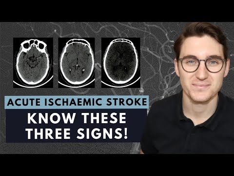
How to read a CT brain scan: Acute ischaemic stroke for beginners
 0:04:57
Stroke: Evolution from acute to chronic infarction - radiology video tutorial (CT, MRI)
 0:00:26
CT Scan of the Brain: Normal or Not? #doctor #CTHead #venicebeach
 0:02:28
Normal Pressure Hydrocephalus CT Scan Brain.
 0:20:32
CT head anatomy for Medical students , residents and clinicians.
 0:00:57
10 Important CT head scans! #neurology #radiology #ctscan
 0:00:38
Normal and Alzheimer's brains dataset comparison.
 0:12:09
Normal CT Scan of brain study
 0:00:11
CT scan Brain 🆚 MRI Brain/Pathology detailing #aiims
 0:00:27
CT and MRI scans can be normal after brain injury
 0:02:32
What’s the Difference Between an MRI and a CT?
 0:00:16
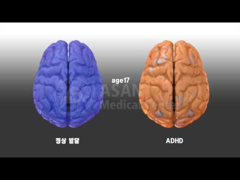
Compare normal vs ADHD brain
 0:00:41
Brain MRI 🧲 🧠 #mri #radiology
 0:00:06
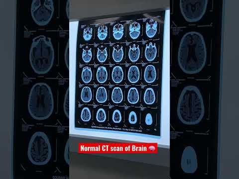
Normal CT scan of Brain 🧠
 0:00:33
CT Scan Of Brain 🧠
 0:00:36
Ct Scan of Child Brain 🧠
 0:05:43
Understand Your Scan: MS MRI and Brain Atrophy
 0:10:22
Multiple Sclerosis MRI
Комментарии