filmov
tv
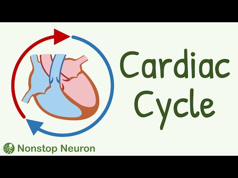
Cardiac cycle, stages, physiology, Diastole and systole in the cardiac cycle.

Показать описание
.
Chapters
0:00 Introduction
0:28 Phases of Cardiac Cycle
2:46 The Systole
The cardiac cycle is the performance of the human heart from the ending of one heartbeat to the beginning of the next. It consists of two periods: one during which the heart muscle relaxes and refills with blood, called diastole, following a period of robust contraction and pumping of blood, dubbed systole. After emptying, the heart immediately relaxes and expands to receive another influx of blood returning from the lungs and other systems of the body, before again contracting to pump blood to the lungs and those systems. A normally performing heart must be fully expanded before it can efficiently pump again. Assuming a healthy heart and a typical rate of 70 to 75 beats per minute, each cardiac cycle, or heartbeat, takes about 0.8 seconds to complete the cycle.[2] There are two atrial and two ventricle chambers of the heart; they are paired as the left heart and the right heart—that is, the left atrium with the left ventricle, the right atrium with the right ventricle—and they work in concert to repeat the cardiac cycle continuously, (see cycle diagram at right margin). At the start of the cycle, during ventricular diastole–early, the heart relaxes and expands while receiving blood into both ventricles through both atria; then, near the end of ventricular diastole–late, the two atria begin to contract (atrial systole), and each atrium pumps blood into the ventricle below it.[3] During ventricular systole the ventricles are contracting and vigorously pulsing (or ejecting) two separated blood supplies from the heart—one to the lungs and one to all other body organs and systems—while the two atria are relaxed (atrial diastole). This precise coordination ensures that blood is efficiently collected and circulated throughout the body.[4]
 0:04:30
Cardiac cycle, stages, physiology, Diastole and systole in the cardiac cycle.
 0:10:13
Cardiac Cycle || Systole, Diastole, Blood flow in heart, Movement of Valves
 0:10:44
Cardiac Cycle | Events | Part 1 | Cardiac Physiology
 0:01:49
The cardiac cycle
 0:23:59
Cardiovascular | Cardiac Cycle
 0:15:18
The Cardiac Cycle - Systole and Diastole - Atria and Ventricles - Physiology and Biology
 0:08:46
Cardiac Cycle
 0:06:30
The Cardiac Cycle
 0:18:50
Cardiovascular | Cardiac Cycle: Digital Version
 0:01:43
The difference between systole and diastole
 0:06:37
The Cardiac Cycle Phase 1 - Atrial Systole Made EASY!!
 0:14:33
A-level- CARDIAC CYCLE. Diastole, atrial systole, ventricular systoles +the pressure +volume changes
 0:06:26
Changes in pressure-volume loops
 1:12:18
Cardiac Cycle | Cardiology | Systole & Diastole | Cardiovascular🫀
 0:04:57
The Cardiac Cycle: Systole & Diastole (Part 1) | Sketchy Medical | USMLE Step 1
 0:00:35
The Cardiac Cycle Made Ridiculously Simple 🫀
 0:04:20
Cardiac Cycle:Part 3-Ventricular Diastole
 0:22:29
Cardiovascular Physiology : Cardiac Cycle and Heart sounds
 0:08:14
Circulatory System and Pathway of Blood Through the Heart
 0:04:16
Depolarization and Repolarization of Heart: Action Potential (Atrial & Ventricular) Animation
 0:06:41
Cardiac Cycle and Conduction System of Heart [Physiology Animation]
 0:01:38
How the cardiac cycle is produced by electrical impulses in the heart
 0:00:12
Cardiac Cycle Physiology - Animation - AUDIO & ECG
 0:08:19
THE CARDIAC CYCLE - Phases, Pressure Changes, ECG/EKG
Комментарии