filmov
tv
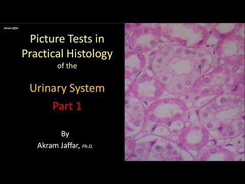
Picture tests in histology of the renal system 1

Показать описание
You may use the video as a revision for the topic or as a self-assessment tool. For the purpose of self-assessment, pause the video at the beginning of each slide and take your time in reading the question and coming up with the answer then replay the video to confirm your answer and listen to further comments and explanations.
00:00 Introduction
00:28 Q1 Glomerular filter
03:47 Q2 Matching proximal and distal convoluted tubules
07:16 Q3 Muscle fibers and epithelium
09:12 Q4 Differentiating the renal cortex from the medulla
11:37 Q5 Identifying the component layers of a tubular structure
13:17 Q6 Identify an organ and describe the location of a similar type of epithelium
15:05 Q7 Deep into the kidney
After completing this video you will be able to:
Differentiate between
-Proximal convoluted tubule, distal convoluted tubule, collecting duct, thick limb of the loop of Henle, thin limb of the loop of Henle, capillary lumen.
-Renal cortex and medulla.
Describe the structural components of the glomerular filter.
Explain the presence of a brush border and dark cytoplasm in proximal convoluted tubules and the abundance of profiles of proximal convoluted tubules in comparison to the distal.
Identify transitional epithelium, mesothelium, urinary bladder, ureter, minor calyx, renal papilla, detrusor muscle, glomerulus, Bowman’s capsule, renal corpuscle, and umbrella (dome-shaped cells).
Presented and edited by Dr. Akram Jaffar, Ph.D.
Subscribe to the channel to receive updates. Feedback is highly appreciated by channel viewers
Related accounts
Some images, with gratitude, were cited in:
 0:15:07
Picture tests in the histology of the gastrointestinal system 1
 0:17:05
Picture tests in histology of the renal system 1
 0:13:07
Picture tests in histology of the cardiovascular system 1
 0:14:50
Picture tests in practical histology of lymphatic tissue part 1
 0:18:51
Picture tests in histology of the respiratory system
 0:15:23
Picture test in histology of the endocrine glands
 0:18:19
Picture Tests in Histology CVS Respiratory Renal
 0:13:38
Picture tests in histology reproductive system - female
 0:17:16
Picture tests in histology of the gastrointestinal system 2
 0:13:58
Picture tests in histology of the renal system 2
 0:12:18
Picture tests in histology of the gastrointestinal system 6
 0:16:46
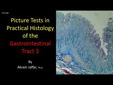
Picture tests in histology of the gastrointestinal system 3
 0:14:26
Picture tests in histology of the cardiovascular system 2
 0:17:30
Picture tests in practical histology of lymphatic tissue part 2
 0:14:50
Picture tests in histology of the gastrointestinal system 5
 0:16:40
Picture tests in histology of the gastrointestinal system 4
 0:13:40
Identifying Epithelium | Review and Practice Questions
 0:25:45
Identifying Tissues | Review and Practice
 0:27:47
Digestive System Histology | Review and Practice
 0:04:11
Identifying Cartilage | Review and Practice Questions
 0:37:43
Introduction to Histology
 0:00:49
Kidney histology explained in 1 minute | 1 minute histology
 0:09:24
Intro to Histology: The Four Tissue Types | Corporis
 0:02:38
Histology | Compact Bone (Osseous Tissue)
Комментарии