filmov
tv
Using microCT to Assess Bone Loss in Preclinical Models

Показать описание
Using microCT to Assess Bone Loss in Preclinical Models
Bone loss is a common medical condition associated with aging, a wide spectrum of diseases, as well as certain treatments (e.g.,corticosteroids). Chronic inflammation, asthma, celiac disease, diabetes, hyperthyroidism, multiple sclerosis, hemophilia, arthritis, periodontal disease, and various cancers and metastatic disease, all manifest bone destruction. With the aging of our population, there is an urgent need for alternative therapies with minimal side effects to treat and/or prevent osteoporotic disease. Ovariectomized or orchidectomized mice or rats are commonly used animal models of osteoporosis to study bone resorption inhibition and bone formation promoting effects. Quantification of1) bone mineral density and 2) bone microstructure morphometric parameters offers invaluable data to further our understanding of the effects of novel treatment on disease progression. Preclinical micro-computed tomography (microCT) offers the researcher a tool for imaging bones in animal models.
PerkinElmer’s Quantum microCT imaging system uniquely delivers a large dynamic range, offering the flexibility to:
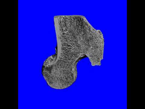
1)imaging bones quantitatively in vivo with imaging protocols featuring radiation doses low enough to facilitate longitudinal studies; as well as 2) imaging bones at the highest resolution ex vivo. Beyond scanning, microCT reconstruction and quantitative analysis software complete the tool package essential for bone research.
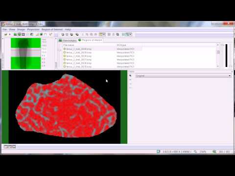
In this webinar, Dr. Alexandra De Lille (Director of Technical Applications for in vivo Imaging at PerkinElmer) will highlight recent research in the field of bone loss. The ability of microCT scanning to monitor disease longitudinally, to detect fine bone architecture change and to differentiate treatment efficacy, will be discussed. However, simply detecting bone loss is not enough. Quantification of bone changes can be time-consuming and user-dependent. PerkinElmer’s AccuCT™ software allows for automated bone identification, segmentation and analysis, thus increasing throughput and accuracy while illuminating advanced bone parameter insight. The Quantum microCT scanner and AccuCT™ advanced bone analysis software are enabling exciting research developments in applications where bone loss is observed. Join us for what is sure to be a thought-provoking discussion on current and future states of microCT imaging and bone loss. Dr. De Lille will also answer your questions following the presentation.
What you will learn:
- Novel methods for quantitative imaging of bone loss in animal models
- The power of low dose, fast, longitudinal in vivo and, high resolution
ex vivo bone imaging with the high-speed QuantumGX microCT system
- How AccuCT advanced bone software can easily and efficiently segment bones and quantitate bone parameters, significantly increasing analysis throughput
Speaker:
Alexandra De Lille
Director of Technical Applications
In Vivo Imaging at PerkinElmer
Original Broadcast Date: September 29th, 2017
Bone loss is a common medical condition associated with aging, a wide spectrum of diseases, as well as certain treatments (e.g.,corticosteroids). Chronic inflammation, asthma, celiac disease, diabetes, hyperthyroidism, multiple sclerosis, hemophilia, arthritis, periodontal disease, and various cancers and metastatic disease, all manifest bone destruction. With the aging of our population, there is an urgent need for alternative therapies with minimal side effects to treat and/or prevent osteoporotic disease. Ovariectomized or orchidectomized mice or rats are commonly used animal models of osteoporosis to study bone resorption inhibition and bone formation promoting effects. Quantification of1) bone mineral density and 2) bone microstructure morphometric parameters offers invaluable data to further our understanding of the effects of novel treatment on disease progression. Preclinical micro-computed tomography (microCT) offers the researcher a tool for imaging bones in animal models.
PerkinElmer’s Quantum microCT imaging system uniquely delivers a large dynamic range, offering the flexibility to:
1)imaging bones quantitatively in vivo with imaging protocols featuring radiation doses low enough to facilitate longitudinal studies; as well as 2) imaging bones at the highest resolution ex vivo. Beyond scanning, microCT reconstruction and quantitative analysis software complete the tool package essential for bone research.
In this webinar, Dr. Alexandra De Lille (Director of Technical Applications for in vivo Imaging at PerkinElmer) will highlight recent research in the field of bone loss. The ability of microCT scanning to monitor disease longitudinally, to detect fine bone architecture change and to differentiate treatment efficacy, will be discussed. However, simply detecting bone loss is not enough. Quantification of bone changes can be time-consuming and user-dependent. PerkinElmer’s AccuCT™ software allows for automated bone identification, segmentation and analysis, thus increasing throughput and accuracy while illuminating advanced bone parameter insight. The Quantum microCT scanner and AccuCT™ advanced bone analysis software are enabling exciting research developments in applications where bone loss is observed. Join us for what is sure to be a thought-provoking discussion on current and future states of microCT imaging and bone loss. Dr. De Lille will also answer your questions following the presentation.
What you will learn:
- Novel methods for quantitative imaging of bone loss in animal models
- The power of low dose, fast, longitudinal in vivo and, high resolution
ex vivo bone imaging with the high-speed QuantumGX microCT system
- How AccuCT advanced bone software can easily and efficiently segment bones and quantitate bone parameters, significantly increasing analysis throughput
Speaker:
Alexandra De Lille
Director of Technical Applications
In Vivo Imaging at PerkinElmer
Original Broadcast Date: September 29th, 2017
 0:37:49
 0:58:21
 0:25:24
 0:00:34
 0:10:53
 0:02:36
 0:47:15
 0:16:33
 0:28:31
 0:05:39
 0:00:07
 0:00:27
 0:16:33
 0:05:23
 0:32:15
 0:05:54
 0:07:34
 0:00:13
 0:25:24
 0:16:35
 0:27:43
 0:05:23
 0:02:49
 0:01:10