filmov
tv
Rough Endoplasmic Reticulum Structure and Function | Rough ER Simplified

Показать описание
Learn more about the structures and functions of the rough endoplasmic reticulum in this quick introduction and overview.
---VIDEOS AND PLAYLISTS---
---DIVE IN---
---MY GEAR---
---RECOMMENDED STUDY GUIDES---
---STUDY RESOURCES---
DISCLAIMER: This video and description contains affiliate links, which means that if you click on some of the product links, I’ll receive a small commission. This helps support the channel and allows us to continue to make videos like this. Thank you for the support!
Music by "Operatic 3" by Vibe Mountain
---Transcript---
Thanks for stopping by, this is 2 minute classroom and today we are talking about the Rough Endoplasmic Reticulum, or Rough ER for short.
I created this channel save you time in your studies so that you can spend more time doing what you enjoy. So consider subscribing and hitting the bell if you want to spend less time studying.
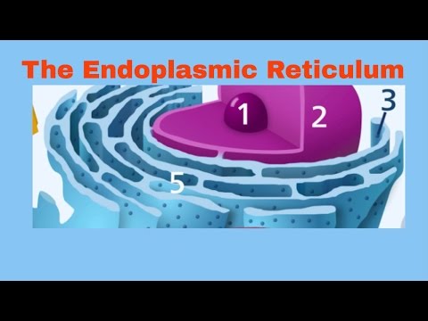
The rough ER is a series of interconnected membranous structures that appears studded under powerful microscopes. These studs are actually ribosomes and give this portion of the ER its rough appearance and name.
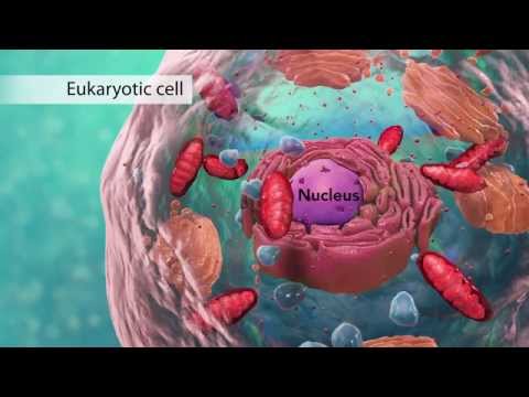
It sits next to the nucleus in the cell and is actually continuous with the outer membrane of the nucleus, the nuclear envelope. The Rough ER is responsible for the synthesis and modification of proteins. You can learn more about proteins by clicking the icon in the upper right hand corner
The proteins that are modified in and pass through the Rough ER are primarily membrane bound proteins that will function in either the cell membrane or the membranes of other organelles within the cell.
Some proteins from the rough ER will be secreted out of the cell in vesicles to perform functions throughout the body, such as signaling between nerve cells.
You can learn more about cells by watching this video, or prepare for an upcoming test with this test tips and strategies playlist. You can also watch this video of mine that YouTube thinks you’ll enjoy.
Thanks for watching, and I’ll catch you next time.
Some images adapted from Wikipedia and OpenStax Biology Text
---VIDEOS AND PLAYLISTS---
---DIVE IN---
---MY GEAR---
---RECOMMENDED STUDY GUIDES---
---STUDY RESOURCES---
DISCLAIMER: This video and description contains affiliate links, which means that if you click on some of the product links, I’ll receive a small commission. This helps support the channel and allows us to continue to make videos like this. Thank you for the support!
Music by "Operatic 3" by Vibe Mountain
---Transcript---
Thanks for stopping by, this is 2 minute classroom and today we are talking about the Rough Endoplasmic Reticulum, or Rough ER for short.
I created this channel save you time in your studies so that you can spend more time doing what you enjoy. So consider subscribing and hitting the bell if you want to spend less time studying.
The rough ER is a series of interconnected membranous structures that appears studded under powerful microscopes. These studs are actually ribosomes and give this portion of the ER its rough appearance and name.
It sits next to the nucleus in the cell and is actually continuous with the outer membrane of the nucleus, the nuclear envelope. The Rough ER is responsible for the synthesis and modification of proteins. You can learn more about proteins by clicking the icon in the upper right hand corner
The proteins that are modified in and pass through the Rough ER are primarily membrane bound proteins that will function in either the cell membrane or the membranes of other organelles within the cell.
Some proteins from the rough ER will be secreted out of the cell in vesicles to perform functions throughout the body, such as signaling between nerve cells.
You can learn more about cells by watching this video, or prepare for an upcoming test with this test tips and strategies playlist. You can also watch this video of mine that YouTube thinks you’ll enjoy.
Thanks for watching, and I’ll catch you next time.
Some images adapted from Wikipedia and OpenStax Biology Text
 0:01:32
Rough Endoplasmic Reticulum Structure and Function | Rough ER Simplified
 0:30:03
Endoplasmic Reticulum Structure & Function
 0:09:06
Endoplasmic reticulum: structure and function
 0:02:35
Endoplasmic Reticulum Rough and Smooth ER
 0:02:08
The Endoplasmic Reticulum - The transportation system of the cell
 0:07:22
Biology: Cell Structure I Nucleus Medical Media
 0:04:37
Endoplasmic Reticulum: Structure & Functions
 0:03:49
Practically's Concepts - Cell Organelles Endoplasmic Reticulum - #LearnPractically
 0:23:05
Nucleus : Master Mind Of Cell - NEET NCERT Biology - Cell Unit Of Life
 0:07:38
Endoplasmic Reticulum structure and functions | Video 6
 0:05:05
Protein Synthesis and the Rough Endoplasmic Reticulum
 0:03:55
Endoplasmic Reticulum: Structure and Functions|| SER|| RER|| Cell biology|| Biology
 0:02:24
Endoplasmic Reticulum Structure and Function : Cell
 0:03:30
Endoplasmic Reticulum in 3 Minutes - (Structure and functions)
 0:11:40
Endoplasmic reticulum and Golgi bodies | Biology | Khan Academy
 0:05:15
Practically's Concepts - Golgi Apparatus - #LearnPractically
 0:08:16
Cell Organelles and Structures Review
 0:08:15
Endoplasmic reticulum | from structure to function | Rough endoplasmic reticulum
 0:02:17
how to draw the diagram of endoplasmic reticulum, er easily,
 0:06:16
The Rough Endoplasmic Reticulum | Structure | Function | Cell Biology | Basic Science Series
 0:03:52
Endoplasmic Reticulum Structure And Functions | Class 11 Biology
 0:16:05
1-5 Nucleus, Endoplasmic Reticulum (Smooth and Rough ER), & Golgi Body (Cambridge A Level Biolog...
 0:01:42
Smooth Endoplasmic Reticulum Function and Structure | Cell Biology Simplified
 0:11:38
Endoplasmic Reticulum
Комментарии