filmov
tv
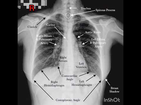
Torso Chest PA x-ray 🩻

Показать описание
Chest
Region
lung, trachea, bronchus, heart, diaphragm, mediastinum, costophrenic angle
Pathology
lung disease, mediastinal disease and heart disease
IR Size
35 × 43 cm (14 × 17 inch)
SID
180 cm (72 inches)
Central Ray
perpendicular beam directed at the center point of the chest, 8~10cm inferior to the jugular notch at the level of 7th thoracic vertebra.
Respiration
suspended with deep inspiration
Position
1. The patient is in an erect position.
2. Place the chin on the vertical image receptor device. Place the hands either on the hip region or on the back of the device with arms surrounding the receptor device, and place the shoulders firmly on the image receptor.
3. Place the midsagittal plane of the body at the center of the IR and make sure the chest is not rotated.
Collimation
Include the entire lung field.
Evaluation
1. Bilateral sternoclavicular joints (S-C joint) should be at the same level with spinal column.
2. Lung apex should be superior to the clavicle.
3. Scapula must not overlap with lungs.
4. Patient's heart and aortic arch must be visible with the diaphragm below the 12th rib.
5. Bilateral lung field must be clearly demonstrated in long scale contrast state.
kVp
100
mAs
4
Tip
1. Larger contrast of subject among surrounding pulmonary blood vessels can be obtained after putting air in Pulmonary Alveoli if the X-ray inspection is implemented holding a breathe when second inspiration is over.
2. Move big breast to lateral sides of lung field.
3. Place image receptor (IR) widthwise for patient with big body shape.
4. In order to reduce density differences between heart shadow and lung shadow, high voltage shooting is recommended.
5. If patient do not have consciousness or cannot move, Chest AP Supine Projection can be used.
#radiology #science #xray #bmw
 0:07:02
 0:04:42
 0:03:28
 0:12:37
 0:00:50
 0:06:46
 0:08:51
 0:00:15
 0:03:07
 0:02:54
 0:00:09
 0:00:15
 0:00:08
 0:00:16
 0:19:44
 0:00:08
 0:10:04
 0:00:19
 0:03:23
 0:00:33
 0:08:07
 0:01:00
 0:01:34
 0:01:00