filmov
tv
Anatomy of The Human Eye Ball | Structure & Function | Cornea & Sclera

Показать описание
#drnajeeb #medicines #medicaleducation #pharmacology #humaneye #eyeanatomy
Anatomy of The Human Eye Ball | Structure & Function | Cornea & Sclera
Like this video?
---------------------------------------------------------------------------------------------------------------------------
Why sign up for premium membership? Here's why!
Membership Features for premium website members.
1. More than 1000+ Medical Lectures.
2. Basic Medical Sciences & Clinical Medicine.
3. Mobile-friendly interface with android and iOS apps.
4. English subtitles and new videos every week.
5. Download option for offline video playback.
6. Fanatic customer support and that's 24/7.
7. Fast video playback option to learn faster.
8. Trusted by over 2M+ students in 190 countries.
---------------------------------------------------------------------------------------------------------------------------
▬▬▬▬▬▬▬▬▬▬ Contents of this video ▬▬▬▬▬▬▬▬▬▬
00:00:00 Introduction to the structure of eyeball
00:00:57 Detailed explanation on coats/Layers of the eyeball
00:17:39 Purpose of the three coats/layers of the eyeball 17:39
00:18:29 What is Lens, vitreous humour and aqueous humour
00:21:00 Review of the basic structure of the eyeball
---------------------------------------------------------------------------------------------------------------------------
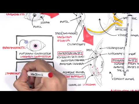
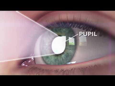
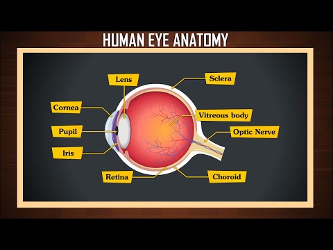
Humans have two eyes, situated on the left and the right of the face. The eyes sit in bony cavities called the orbits, in the skull. There are six extraocular muscles that control eye movements. The front visible part of the eye is made up of the whitish sclera, a colored iris, and the pupil. A thin layer called the conjunctiva sits on top of this. The front part is also called the anterior segment of the eye.
The eye is not shaped like a perfect sphere, rather it is a fused two-piece unit, composed of an anterior (front) segment and the posterior (back) segment. The anterior segment is made up of the cornea, iris, and lens. The cornea is transparent and more curved and is linked to the larger posterior segment, composed of the vitreous, retina, choroid, and the outer white shell called the sclera. The cornea is typically about 11.5 mm (0.45 in) in diameter, and 0.5 mm (500 μm) in thickness near its center. The posterior chamber constitutes the remaining five-sixths; its diameter is typically about 24 mm (0.94 in). The cornea and sclera are connected by an area termed the limbus. The iris is the pigmented circular structure concentrically surrounding the center of the eye, the pupil, which appears to be black. The size of the pupil, which controls the amount of light entering the eye, is adjusted by the iris' dilator and sphincter muscles.
Light energy enters the eye through the cornea, through the pupil, and then through the lens. The lens shape is changed for near focus (accommodation) and is controlled by the ciliary muscle. Photons of light falling on the light-sensitive cells of the retina (photoreceptor cones and rods) are converted into electrical signals that are transmitted to the brain by the optic nerve and interpreted as sight and vision.
---------------------------------------------------------------------------------------------------------------------------
Join this channel to get access to perks:
Anatomy of The Human Eye Ball | Structure & Function | Cornea & Sclera
Like this video?
---------------------------------------------------------------------------------------------------------------------------
Why sign up for premium membership? Here's why!
Membership Features for premium website members.
1. More than 1000+ Medical Lectures.
2. Basic Medical Sciences & Clinical Medicine.
3. Mobile-friendly interface with android and iOS apps.
4. English subtitles and new videos every week.
5. Download option for offline video playback.
6. Fanatic customer support and that's 24/7.
7. Fast video playback option to learn faster.
8. Trusted by over 2M+ students in 190 countries.
---------------------------------------------------------------------------------------------------------------------------
▬▬▬▬▬▬▬▬▬▬ Contents of this video ▬▬▬▬▬▬▬▬▬▬
00:00:00 Introduction to the structure of eyeball
00:00:57 Detailed explanation on coats/Layers of the eyeball
00:17:39 Purpose of the three coats/layers of the eyeball 17:39
00:18:29 What is Lens, vitreous humour and aqueous humour
00:21:00 Review of the basic structure of the eyeball
---------------------------------------------------------------------------------------------------------------------------
Humans have two eyes, situated on the left and the right of the face. The eyes sit in bony cavities called the orbits, in the skull. There are six extraocular muscles that control eye movements. The front visible part of the eye is made up of the whitish sclera, a colored iris, and the pupil. A thin layer called the conjunctiva sits on top of this. The front part is also called the anterior segment of the eye.
The eye is not shaped like a perfect sphere, rather it is a fused two-piece unit, composed of an anterior (front) segment and the posterior (back) segment. The anterior segment is made up of the cornea, iris, and lens. The cornea is transparent and more curved and is linked to the larger posterior segment, composed of the vitreous, retina, choroid, and the outer white shell called the sclera. The cornea is typically about 11.5 mm (0.45 in) in diameter, and 0.5 mm (500 μm) in thickness near its center. The posterior chamber constitutes the remaining five-sixths; its diameter is typically about 24 mm (0.94 in). The cornea and sclera are connected by an area termed the limbus. The iris is the pigmented circular structure concentrically surrounding the center of the eye, the pupil, which appears to be black. The size of the pupil, which controls the amount of light entering the eye, is adjusted by the iris' dilator and sphincter muscles.
Light energy enters the eye through the cornea, through the pupil, and then through the lens. The lens shape is changed for near focus (accommodation) and is controlled by the ciliary muscle. Photons of light falling on the light-sensitive cells of the retina (photoreceptor cones and rods) are converted into electrical signals that are transmitted to the brain by the optic nerve and interpreted as sight and vision.
---------------------------------------------------------------------------------------------------------------------------
Join this channel to get access to perks:
 0:09:55
Eyeball Anatomy
 0:11:25
Anatomy - Eye Overview
 0:02:49
EYE ANATOMY IN 3 MINUTES!
 0:23:21
Anatomy of The Human Eye Ball | Structure & Function | Cornea & Sclera
 0:17:05
Basic Eye Anatomy and Physiology
 0:07:20
Eye Anatomy and Function - Made Easy
 0:03:28
Eyeball: structure and function (preview) - Human Anatomy | Kenhub
 0:10:22
Parts of the eye | Human eye & the colourful world | Khan Academy
 0:02:42
'What is the Eye Made Of? A Journey Inside Our Vision'
 0:45:46
Special Senses | Eye Anatomy
 0:01:01
How eyes works? (Animation) explained within one minute.
 0:14:20
The Coolest Eyeball Video You'll Ever See
 0:01:45
Human A&P: Anatomy of the Eye
 0:11:17
Why the Eye Has the Fastest Muscles in the Human Body
 0:04:42
Human Anatomy Eye
 0:03:04
Human Eye Anatomy | Structure and function | Parts of the Eye
 0:09:21
Anatomy of the Eye
 0:04:17
Human Eye - The Dr. Binocs Show | Best Learning Videos For Kids | Peekaboo Kidz
 0:10:09
Eye Anatomy | Review and Practice
 0:09:39
Vision: Crash Course Anatomy & Physiology #18
 0:08:22
What Happens Inside Your Eyes - 3D Animation
 0:09:50
Structure of Human Eye | Photoreceptor | Biology lecture
 0:20:38
Structure of human Eye: Human Eye Anatomy
 0:17:04
Structure of Human Eye | Anatomy of Eyeball | Eye Anatomy
Комментарии